Visual cortex
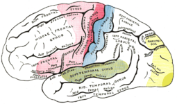
The visual cortex (also visual cortex ) is that part of the cerebral cortex , of the visual system is one which, in turn, the visual perception possible. The visual cortex occupies most of the occipital lobe of the brain. According to Korbinian Brodmann's brain map, areas 17, 18 and 19 correspond to it. It is divided into the primary visual cortex (V1) and secondary or tertiary (associative, V2 – V5) areas. Histologically , it is characterized in primates by the high cell density and the comparatively small thickness (in humans 1.5–2 mm). Area 17 (also area striata ) directly represents the opposite half of the field of vision and is built up “retinotop”, which means that points shown next to each other on the retina are also next to each other here. The cells that represent the fovea are clearly overrepresented and take up about half of the area striata.

| Primary motor area | |
| Pre / supplementary motor areas | |
| Primarily sensitive areas | |
| Sensitive areas of association | |
| Fields of view | |
| Audio fields |
Primary visual cortex (V1)
histology
The primary visual cortex ( Brodmann area 17, V1; area striata ), like the entire neocortex (differentiated from archi- and palaeocortex), is divided into six layers:
- Layer I: molecular layer
- Layer II: outer granular layer
- Layer III: outer pyramidal layer
- Layer IV: inner granular layer
- Layer V: inner pyramidal cell layer
- Layer VI: multiformed layer.
In addition to the layer organization, there is a columnar (columnar) structure in V1, as in the rest of the cortex, which in each case reflects an orientation.
Afferents and internal organization
The primary visual cortex area V1 has a particularly thick layer IV. Many afferent fibers of the neurons of the corpus geniculatum laterale (CGL) end here. Layer IV is further subdivided into A, B and C, C in turn into Cα and Cβ. The axons of magnocellular layers 1 and 2 of the CGL end in layer IVCα. These neurons, in turn, are derived from ganglion cells of the retina with large receptive fields that have a high temporal but low spatial resolution. The work of David Hubel and Torsten Wiesel revealed that neurons of the IVCα layer are orientation-selective. Individual groups of these cells each respond to visual stimuli along the center of their elongated receptive fields up to a deviation of 10 °. These cells are called simple cells . They project into slice IVB, where they are interconnected binocularly and have directional selectivity (M channel).
Parvocellular neurons of layers III – VI of the CGL end in layer IVCβ and, to a lesser extent, in layers IVA and I. They receive afferents of the ganglion cells with small receptive fields (fovea), show color sensitivity and have a high spatial resolution.
Further cortical areas receive the information about movements in the visual field from the inputs of the layer IVCα. From the inputs of the IVCβ layer, other cortical areas receive information about the shape and sometimes the color of objects in the visual field.
Lesions of magnocellular layers in monkeys caused them to lose the ability to perceive movement, whereas damage to the parvocellular layers prevented the perception of color, fine textures as well as shapes and spatial depth.
Layer IV has ocular dominance bands, also known as ocular dominance columns , which can be represented by retrograde axonal transport of radioactive proline (or other amino acids ).
Secondary and tertiary visual cortex (V2 – V5)
The V2 is located according to the areas of Korbinian Brodmann in area 18 and has a stripe structure that can be made visible by histological staining of the cytochrome oxidase activity. According to Livingstone and Hubel (1988), these stripes are associated with the processing of individual aspects of perception, form, perception and stereo vision and then have efferents in the higher brain areas.
After the interconnection in V1 and V2, the processing is divided into a dorsal path through the parietal lobe , into the motor areas along the central groove and a ventral path that leads into the areas of the temporal lobe , to which the beginning of object recognition is ascribed. Among other things, V4 and the inferotemporal cortex are assigned to the ventral path. The dorsal path runs through V5 (also called the mediotemporal cortex). The association cortices are, however, not as precisely mapped as z. B. V1, as there are inter-individual differences.
See also
literature
- Otto Detlev Creutzfeldt : Cortex cerebri. Springer (1983). ISBN 3-540-12193-5
- Karl Zilles , G. Rehkämper: Functional Neuroanatomy. Springer, Berlin 1993. ISBN 3-540-54690-1
- D. Drenckhahn, W. Zenker: Benninghoff. Anatomy. Urban & Schwarzenberg, Munich 1994 ISBN 3-541-00255-7
- Eric Kandel , James Schwartz, Thomas Jessell : Neurosciences. Spectrum Academic Publishing House, Heidelberg 1996 ISBN 3-86025-391-3
- Edwin Clarke, CD O'Mally: The Human Brain and Spinal Cord: A Historical Study Illustrated by Writings from Antiquity to the 20th Century , University of California Press, 1968
- Mitchell Glickstein, Giacomo Rizzolattic: Francesco Gennari and the structure of the cerebral cortex , Trends in Neurosciences, Volume 7, Issue 12, pp. 464-467, December 1, 1984, doi : 10.1016 / S0166-2236 (84) 80255-6
- Mitchell Glickstein, David Whitteridge: Tatsuji Inouye and the mapping of the visual fields on the human cerebral cortex , Trends in Neurosciences, Volume 10, Issue 9, pp. 350–353, January 1, 1987, doi : 10.1016 / 0166-2236 ( 87) 90066-X