Visual system

The visual system is that part of a nervous system that deals with processing visual information. The visual system comprises the eye with retina (retina), the optic nerve , parts of the thalamus and the brain stem, and the visual cortex .
Dioptric apparatus
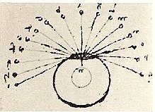
The dioptric apparatus of the eye ( cornea , aqueous humor , lens , vitreous humor ) creates an upside-down and reversed image of the surroundings in the field of vision on the retina .
The fovea is the point with the greatest possible sharpness of the image. However, the eye can only remain on such a fixation point for around 0.2 to 0.3 seconds ( optokinetic nystagmus ).
Around half of the optic nerve (below in yellow) goes from the fovea to the visual center. The remaining half of the optic nerve is reserved for the peripheral system, which captures up to 90 compressed images of the field of view per second.
Visual pathway

The light stimuli are registered by the sensory cells of the retina, the rods and cones , and displayed as changes in their membrane potential ( receptor potential ). Action potentials can be triggered here via the bipolar cells passed on to the ganglion cells . The extensions of the retinal ganglion cells form the 2nd cranial nerve (optic nerve) , which forwards the action potentials .
The visual pathway conducts the action potentials to the visual cortex: After entering the cranial cavity , the optic nerves of both eyes cross at the junction of the optic nerves ( optic chiasm ). The outer (temporal) fibers continue to run uncrossed, while the inner (nasal) fibers cross to the opposite side. In this way, the fibers of the left half of the retina in both eyes run into the left half of the brain and those of the right half of the retina into the right. In the two optic tracts these nerve fibers run to the Lateral geniculate lateral geniculate nucleus of the thalamus, from where they have the broad optic radiation ( optic radiation ) to the visual cortex ( visual cortex are forwarded).
Visual perception
Visual perception means - going beyond pure "seeing" - an "explicit symbolic description of the observed scene". For this purpose, the image of the scene projected on the retina by the optical apparatus is already analyzed in the retina (brightness, color, contrasts, movement) and edited (brightness compensation, contrast enhancement). When it is transmitted via the optic nerves and the knees, the spatial relationships of the receptors in the relationships of the nerve tracts and synapses are preserved (so-called retinotopy ). This positional relationship can be demonstrated in the visual cortex as a neural map. The activity of the nerve cells on this map represents the perception of the scene, but in a distorted form: the left and right halves are separated from each other in the right and left hemispheres, the center of the scene (fixation point) as the most important part of the scene is represented by a larger region than the edge.
The recognition of individual elements and their meaning is probably carried out by comparing them with already stored experiences (scenes linked to body feeling, emotions, smell, noises and much more).
Eye reflexes
The nerve tracts of the eye reflexes are linked to the visual pathway. Reflexes that control the automatic fixation of static or moving objects use the spatial information from the visual cortex that has already been processed.
Protective reflexes
- Pupillary reflex - compensation of abrupt light-dark changes by changing the opening width of the pupil .
- Lid closing reflex to protect against glaring light, drying drafts and foreign bodies.
- Bell's phenomenon: Rotation of the eyes outwards and upwards as part of the blink reflex
- Westphal-Piltz phenomenon, pupil constriction as part of the eyelid-closing reflex
Fixation reflexes
- Accommodation : Adaptation of the optical properties of the lenses to the distance of the fixed object by changing the curvature of the lens
- Convergence : Alignment of the facial lines of both eyes with the distance of the focused object. With objects that are far away, the axes are aligned parallel. The closer the object, the more the eyes have to be turned inward. This is done at the same time by the eye muscle ( rectus medialis muscle ) lying nasally .
- Convergence miosis Pupillary constriction with near fixation
- Follow-up movement , optokinetic nystagmus , vestibulo-ocular reflex and doll's head phenomenon : alignment of the visual axes to compensate for head movements or when following moving objects.
- Saccadic eye movements (saccades) are rapid, jerky eye movements. They serve to supplement the peripheral perception and the already existing ideas. They also occur while dreaming and imagining visual presentations .
- Micro saccade of rapid eye target movements to avoid local adaptation
credentials
- ↑ Hans-Werner Hunziker: In the eye of the reader. Transmedia, Zurich 2006, ISBN 978-3-7266-0068-6 (Original title: In the Eye of the Reader. Foveal and Peripheral Perception. From Letter Recognition to the Joy of Reading. ).
- ↑ Quaderni d'anatomia IV fol. 12 verso, quoted in Sandro Piantanida, Costantino Baroni (ed.), Kurt Karl Eberlein (translation): Leonardo da Vinci - The image of a genius . Documentation of the Leonardo da Vinci exhibition in Milan in 1938. Lüttke-Verlag Berlin undated (1939/40). Reprint Emil Vollmer Verlag 1955. p. 430 limited preview in the Google book search
- ↑ John P. Frisby: Optical Illusions. Seeing, perceiving, memory. Weltbild, Augsburg 1989 ISBN 3-926187-24-7 , p. 182 f.
- ↑ Werner Kahle et al. a .: nervous system and sense organs. dtv, Munich 1978, ISBN 3-423-03019-4 ( dtv Atlas of Anatomy. Volume 3), p. 312 f.
literature
- Semir Zeki : The spiritual image of the world. In: Spectrum of Science. November 1992
- Werner Kahle u. a .: nervous system and sense organs. dtv, Munich 1978, ISBN 3-423-03019-4 ( dtv Atlas of Anatomy. Volume 3), p. 308 f.

