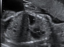
Diaphragmatic hernia
With a diaphragmatic hernia, abdominal organs are shifted into the chest cavity through a weak point or gap in the diaphragm .
to form
There are congenital and acquired diaphragmatic hernias. The congenital forms are malformations . Abdominal organs ( stomach , intestines , liver , spleen ) can enter the chest through a gap in the diaphragm . Sometimes they are associated with other malformations.
In the case of an acquired diaphragmatic hernia, the stomach usually reaches the top of the chest next to the esophagus; with these hiatal hernias, the contents of the abdominal cavity push through the slit-shaped diaphragmatic opening through which the esophagus ( hiatus oesophageus ) runs.
If the upwardly displaced organs are covered by peritoneum - i.e. if there is a so-called hernial sac - one speaks of a real hernia . In congenital hernias there is often no hernial sac.
Congenital diaphragmatic hernia
| Classification according to ICD-10 | |
|---|---|
| Q79.0 | Congenital diaphragmatic hernia |
| Q79.1 | Absence of the diaphragm |
| ICD-10 online (WHO version 2019) | |
frequency
Congenital diaphragmatic hernia: 2–5 in 10,000 births. In 80–90% of cases the gap is on the left.
Cause and consequences
Disturbed development of the diaphragm : The diaphragm develops in the 8th to 10th week of pregnancy. If there is a developmental disorder that creates a gap in the diaphragm, abdominal organs slide into the chest as a result. If the diaphragmatic hernia is on the right, these are mostly parts of the liver and intestines, on the left side it is usually the stomach, intestines, spleen and possibly parts of the liver. Depending on how much space these organs then take up in the chest, the development of the lungs is restricted. On the one hand, this results in a reduced growth of the lungs and, often in addition, a displacement of the heart . In the lung tissue one also often finds a vascular malformation that leads to circulatory problems after birth.
Diagnosis
The congenital diaphragmatic hernia can be shown on ultrasound before birth from the 18th week of pregnancy . If such a diagnosis is made prenatally, further examinations (e.g. magnetic resonance imaging) should be carried out in a specialized center in order to be able to assess the extent of prenatal lung damage, which significantly influences the prognosis for the survival of the children and the desired first aid.
After birth, the children are noticeable because of shortness of breath or guttural belly , an X-ray of the chest then shows that there are other organs in the chest besides the lungs and heart.
therapy
If the diaphragmatic hernia is found before delivery, delivery should be planned in consultation with a specialized center. In some selected cases, in utero therapy will be suggested, in which a balloon is placed in the windpipe for a certain period of time, which is then removed again before delivery.
Otherwise, delivery should take place by means of a planned caesarean section in a center with an attached neonatal intensive care unit and pediatric surgery . After birth, the child is immediately intubated, ventilated and fed with a nasogastric tube to prevent air from entering the parts of the gastrointestinal tract that are in the chest. Otherwise these would require even more space due to the air introduced, which would press on the lungs and further hinder breathing. After the initial treatment, the patient is transferred to the neonatal intensive care unit. The child is first stabilized there and looked for further malformations.
If there is very poor lung function, the use of ECMO (extracorporeal membrane oxygenation) may be necessary to ensure the survival of the children.
The corrective operation is only planned and carried out after the child has been sufficiently stabilized. It should be noted that many children go through a so-called "honeymoon phase" shortly after birth, during which they feel relatively well for a few hours, but then have major problems with their breathing and circulation. The operation can only be planned after further stabilization.
During the operation, the organs are moved back from the chest into the abdomen and the gap in the diaphragm is closed. If the gap is so large that the diaphragm muscles cannot be sewn together, either foreign material is sewn in or part of the muscles of the abdominal wall is used as a replacement for the diaphragm.
When pushing the abdominal organs back into the abdominal cavity, it may happen that there is not enough space in it. In such a case, the abdominal cavity is also closed with foreign material, which is later removed.
forecast
The survival rate varies between 70 and 90% depending on the center.
If the gap in the diaphragm is large, further operations may be necessary, e.g. B. the exchange of foreign material in the course of growth.
Acquired diaphragmatic hernia
In addition to congenital hernias, in which the hernial sac is already formed at birth or in the uterus, hernias can also arise later in life due to pre-existing gaps. If the hernia is the esophageal hiatus , the hernia is called the hiatal hernia . This is the most common form of acquired diaphragmatic hernia.
However, if the break occurs through other gaps, these are eponymous (in the case of Morgagni, Bochdalek or Larrey hernias).
Sources and web links
- ^ Rüdiger Döhler , Günther Heinemann: Eventration of the diaphragm with ipsilateral secondary lung and intestinal nonrotation . Zeitschrift für Kinderchirurgie 25 (1978), pp 258-262
- ↑ a b guideline 006-087l-S1 diaphragmatic hernia diaphragmatic defect
- ↑ UMM: Diaphragmatic hernia: University Hospital Mannheim. Retrieved August 1, 2018 .


