Flagellum: Difference between revisions
m Reverted edits by 189.234.90.110 (talk) to last version by 92.21.92.172 |
No edit summary |
||
| Line 1: | Line 1: | ||
| ⚫ | they reallly suck dick) is a tail-like structure that projects from the [[cell body]] of certain [[prokaryotic]] and [[eukaryotic]] cells, and it functions in [[locomotion]].<ref name="pmid12624192">{{cite journal | author = Bardy SL, Ng SY, Jarrell KF | title = Prokaryotic motility structures | journal = Microbiology (Reading, Engl.) | volume = 149 | issue = Pt 2 | pages = 295–304 | year = 2003 | month = February | pmid = 12624192 | doi = 10.1099/mic.0.25948-0 | url = }}</ref><ref name="Lefebvre_2001">{{cite journal | author = Lefebvre PA | title = Assembly and Motility of Eukaryotic Cilia and Flagella. Lessons from Chlamydomonas reinhardtii | journal = Plant Physiol. | volume = 127 | issue = 4 | pages = 1500–1507 | year = 2001 | month = | pmid = 11743094| doi = 10.1104/pp.010807 | url = }}</ref> An example of a eukaryotic flagellated cell is the [[sperm]] cell, which uses its flagellum to propel itself toward and through the female reproductive tract.<ref name="pmid17148374">{{cite journal | author = Malo AF, Gomendio M, Garde J, Lang-Lenton B, Soler AJ, Roldan ER | title = Sperm design and sperm function | journal = Biol. Lett. | volume = 2 | issue = 2 | pages = 246–9 | year = 2006 | month = June | pmid = 17148374 | doi = 10.1098/rsbl.2006.0449 | url = }}</ref> An example of a flagellated bacterium is the ulcer-causing ''[[Helicobacter pylori]]'', which uses its multiple flagella to propel itself through the mucus lining to reach the stomach [[epithelium]].<ref name="pmid11584108">{{cite journal | author = Lacy BE, Rosemore J | title = Helicobacter pylori: ulcers and more: the beginning of an era | journal = J. Nutr. | volume = 131 | issue = 10 | pages = 2789S–2793S | year = 2001 | month = October | pmid = 11584108 | doi = | url = http://jn.nutrition.org/cgi/content/abstract/131/10/2789S | issn = | format = abstract page }}</ref> Prokaryotic and eukaryotic flagella have some notable differences, such as protein composition, structure, and mechanism of propulsion. Flagella are structurally identical to eukaryotic [[cilia]], although distinctions are sometimes made according to function and/or length.<ref name="pmid6459327">{{cite journal | author = Haimo LT, Rosenbaum JL | title = Cilia, flagella, and microtubules | journal = J. Cell Biol. | volume = 91 | issue = 3 Pt 2 | pages = 125s–130s | year = 1981 | month = December | pmid = 6459327 | doi = 10.1083/jcb.91.3.125s | url = }}</ref> Flagella are cellular ''structures'', not [[organelles]].{{Fact|date=July 2008}} |
||
{{For|the insect anatomical structure|Antenna (biology)}} |
|||
[[Image:Ecoli_flagellum.jpg|thumb|350px|Escherichia coli cells use long, thin structures called flagella to propel themselves. These flagella form bundles that rotate counter-clockwise, creating a torque that causes the bacterium to rotate clockwise.]] |
|||
| ⚫ | |||
The word [[Wiktionary:flagellum|''flagellum'']] comes from the [[Latin]] for [[whip]]. |
The word [[Wiktionary:flagellum|''flagellum'']] comes from the [[Latin]] for [[whip]]. |
||
Revision as of 22:35, 11 October 2008
they reallly suck dick) is a tail-like structure that projects from the cell body of certain prokaryotic and eukaryotic cells, and it functions in locomotion.[1][2] An example of a eukaryotic flagellated cell is the sperm cell, which uses its flagellum to propel itself toward and through the female reproductive tract.[3] An example of a flagellated bacterium is the ulcer-causing Helicobacter pylori, which uses its multiple flagella to propel itself through the mucus lining to reach the stomach epithelium.[4] Prokaryotic and eukaryotic flagella have some notable differences, such as protein composition, structure, and mechanism of propulsion. Flagella are structurally identical to eukaryotic cilia, although distinctions are sometimes made according to function and/or length.[5] Flagella are cellular structures, not organelles.[citation needed]
The word flagellum comes from the Latin for whip.
Types
Three quite distinct types of flagella have so far been distinguished; bacterial, archaeal and eukaryotic.
The main differences among these three types are summarized below:
- Bacterial flagella are helical filaments that rotate like screws.[6][7][8] They provide two of several kinds of bacterial motility.[9][10]
- Archaeal flagella are superficially similar to bacterial flagella, but are different in many details and considered non-homologous.[11][12]
- Eukaryotic flagella - those of animal, plant, and protist cells - are complex cellular projections that lash back and forth.
Sometimes eukaryotic flagella are called cilia or undulipodia to emphasize their distinctiveness.
Bacterial


The bacterial flagellum is made up of the protein flagellin. Its shape is a 20 nanometer-thick hollow tube. It is helical and has a sharp bend just outside the outer membrane; this "hook" allows the helix to point directly away from the cell. A shaft runs between the hook and the basal body, passing through protein rings in the cell's membrane that act as bearings. Gram-positive organisms have 2 of these basal body rings, one in the peptidoglycan layer and one in the plasma membrane. Gram-negative organisms have 4 such rings: the L ring associates with the lipopolysaccharides, the P ring associates with peptidoglycan layer, the M ring is embedded in the plasma membrane, and the S ring is directly attached to the plasma membrane. The filament ends with a capping protein.[13][14]
The bacterial flagellum is driven by a rotary engine made up of protein (Mot complex), located at the flagellum's anchor point on the inner cell membrane. The engine is powered by proton motive force, i.e., by the flow of protons (hydrogen ions) across the bacterial cell membrane due to a concentration gradient set up by the cell's metabolism (in Vibrio species there are two kinds of flagella, lateral and polar, and some are driven by a sodium ion pump rather than a proton pump[15]). The rotor transports protons across the membrane, and is turned in the process. The rotor alone can operate at 6,000 to 17,000 rpm, but with the flagellar filament attached usually only reaches 200 to 1000 rpm.
Flagella do not rotate at a constant speed but instead can increase or decrease their rotational speed in relation to the strength of the proton motive force. Flagellar rotation can move bacteria through liquid media at speed of up to 60 cell lengths/second (sec). Although this is only about 0.00017 km/h, when comparing this speed with that of higher organisms in terms of number of lengths moved per second, it is extremely fast. The fastest land animal, the cheetah, moves at a maximum rate of about 110 km/h, but this represents only about 25 body lengths/sec. Thus, when size is accounted for, prokaryotic cells swimming at 50-60 lengths/sec are actually much faster than larger organisms.[citation needed]
The components of the bacterial flagellum are capable of self-assembly without the aid of enzymes or other factors. Both the basal body and the filament have a hollow core, through which the component proteins of the flagellum are able to move into their respective positions. During assembly, protein components are added at the flagellar tip rather than at the base.[citation needed]
The basal body has several traits in common with some types of secretory pores, such as the hollow rod-like "plug" in their centers extending out through the plasma membrane. Given the structural similarities, it was thought that bacterial flagella may have evolved from such pores; however, it is now known that these pores are derived from flagella.[citation needed]
Flagella arrangement schemes
Different species of bacteria have different numbers and arrangements of flagella. Monotrichous bacteria have a single flagellum (e.g., Vibrio cholerae). Lophotrichous bacteria have multiple flagella located at the same spot on the bacteria's surfaces which act in concert to drive the bacteria in a single direction. In many cases, the bases of multiple flagella are surrounded a specialized region of the cell membrane; the so-called polar membrane. Amphitrichous bacteria have a single flagellum on each of two opposite ends (only one flagellum operates at a time, allowing the bacteria to reverse course rapidly by switching which flagellum is active). Peritrichous bacteria have flagella projecting in all directions (e.g., Escherichia coli).
In some bacteria, such as the larger forms of Selenomonas, the individual flagella are organized outside the cell body, helically twining about each other to form a thick structure called a "fascicle". Other bacteria, such as Spirochetes, have a specialized type of flagellum called an "axial filament" that is located in the periplasmic space, the rotation of which causes the entire bacterium to move forward in a corkscrew-like motion.
Counterclockwise rotation of monotrichous polar flagella thrust the cell forward with the flagella trailing behind. Periodically, the direction of rotation is briefly reversed, causing what is known as a "tumble" in which the cell seems to thrash about in place. This results in the reorientation of the cell. When moving in a favorable direction, "tumbles" are unlikely; however, when the cell's direction of motion is unfavorable (e.g., away from a chemical attractant), a tumble may occur, with the chance that the cell will be thus reoriented in the correct direction.
In some Vibrio (particularly Vibrio parahemolyticus[16]) and related proteobacteria such as Aeromonas, two flagellar systems co-exist, using different sets of genes and different ion gradients for energy. The polar flagella are constitutively expressed and provide motility in bulk fluid, while the lateral flagella are expressed when the polar flagella meets too much resistance to turn.[17][18][19][20][21][22] These provide swarming motility on surfaces or in viscous fluids.
Archaeal
The archaeal flagellum is superficially similar to the bacterial (or eubacterial) flagellum; in the 1980s they were thought to be homologous on the basis of gross morphology and behavior.[23] Both flagella consist of filaments extending outside of the cell, and rotate to propel the cell.
However, discoveries in the 1990s revealed numerous detailed differences between the archaeal and bacterial flagella; these include:
- Bacterial flagella are motorized by a flow of H+ ions (or occasionally Na+ ions); archaeal flagella are almost certainly powered by ATP. The torque-generating motor that powers rotation of the archaeal flagellum has not been identified.
- While bacterial cells often have many flagellar filaments, each of which rotates independently, the archaeal flagellum is composed of a bundle of many filaments that rotate as a single assembly.
- Bacterial flagella grow by the addition of flagellin subunits at the tip; archaeal flagella grow by the addition of subunits to the base.
- Bacterial flagella are thicker than archaeal flagella, and the bacterial filament has a large enough hollow "tube" inside that the flagellin subunits can flow up the inside of the filament and get added at the tip; the archaeal flagellum is too thin to allow this.
- Many components of bacterial flagella share sequence similarity to components of the type III secretion systems, but the components of bacterial and archaeal flagella share no sequence similarity. Instead, some components of archaeal flagella share sequence and morphological similarity with components of type IV pili, which are assembled through the action of type II secretion systems (the nomenclature of pili and protein secretion systems is not consistent).
These differences mean that the bacterial and archaeal flagella are a classic case of biological analogy, or convergent evolution, rather than homology. However, in comparison to the decades of well-publicized study of bacterial flagella (e.g. by Berg), archaeal flagella have only recently begun to get serious scientific attention. Therefore, many assume erroneously that there is only one basic kind of prokaryotic flagellum, and that archaeal flagella are homologous to it. For example, Cavalier-Smith (2002)[23] is aware of the differences between archaeal and bacterial flagellins, but retains the misconception that the basal bodies are homologous.[citation needed]
Eukaryotic


The eukaryotic flagellum is completely different from the prokaryote flagellum in both structure and evolutionary origin. The only shared characteristics among bacterial, archaeal, and eukaryotic flagella are their superficial appearance; they are intracellular extensions used in creating movement. Along with cilia, flagella make up a group of organelles known as undulipodia.
Structure
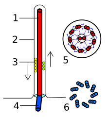
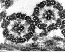
A eukaryotic flagellum is a bundle of nine fused pairs of microtubule doublets surrounding two central single microtubules. The so-called "9+2" structure is characteristic of the core of the eukaryotic flagellum called an axoneme. At the base of a eukaryotic flagellum is a basal body, "blepharoplast" or kinetosome, which is the microtubule organizing center (MTOC) for flagellar microtubules and is about 500 nanometers long. Basal bodies are structurally identical to centrioles. The flagellum is encased within the cell's plasma membrane, so that the interior of the flagellum is accessible to the cell's cytoplasm.
Mechanism
Each of the outer 9 doublet microtubules extends a pair of dynein arms (an "inner" and an "outer" arm) to the adjacent microtubule; these dynein arms are responsible for flagellar beating, as the force produced by the arms causes the microtubule doublets to slide against each other and the flagellum as a whole to bend. These dynein arms produce force through ATP hydrolysis. The flagellar axoneme also contains radial spokes, polypeptide complexes extending from each of the outer 9 microtubule doublets towards the central pair, with the "head" of the spoke facing inwards. The radial spoke is thought to be involved in the regulation of flagellar motion, although its exact function and method of action are not yet understood.
Flagella vs Cilia

Though eukaryotic flagella and cilia are ultrastructurally identical, the beating pattern of the two organelles can be different. In the case of flagella (e.g. the tail of a sperm) the motion is propeller-like. In contrast, beating of cilia consists of coordinated back-and-forth cycling of many cilia on the cell surface. Thus, motile flagella serve for the propulsion of single cells (e.g. swimming of protozoa and spermatozoa), and cilia for the transport of fluids (e.g. transport of mucus by stationary ciliated cells in the trachea). However, cilia are also used for locomotion (through liquids) in organisms such as Paramecium.
Intraflagellar Transport
Intraflagellar transport (IFT), the process by which axonemal subunits, transmembrane receptors, and other proteins are moved up and down the length of the flagellum, is essential for proper functioning of the flagellum, in both motility and signal transduction.[24]
For information on biologists' ideas about how the various flagella may have evolved, see evolution of flagella.
Irreducible complexity
In his 1996 book Darwin's Black Box, intelligent design proponent Michael Behe cited the bacterial flagellum as an example of an irreducibly complex structure that could not have evolved through naturalistic means. Behe argued that the flagellum becomes useless if any one of its constituent parts is removed, and thus could not have arisen through numerous, successive, slight modifications; therefore, it is hopelessly improbable that the proteins making up the flagellar motor could have come together all at once, by chance.[25]
While Behe discussed the immune system and the blood clotting cascade in greater detail, the bacterial flagellum has become a "poster child" for intelligent design proponents and other creationists. It is one of two identified rotary structures found in nature (the other being ATP synthase)[26] and it is billions of years older than Behe's other two examples, which exist in many homologous forms, simplifying the explanation of their origin.[27]
Viable evolutionary pathways have since been proposed for the bacterial flagellum.[28][29][30]
In addition, the Type III secretory system, a molecular syringe which bacteria use to inject toxins into other cells, appears to be a simplified sub-set of the bacterial flagellum's components, meaning that it is much less likely to be irreducibly complex.[31][32]
Behe's arguments have been examined and rejected by the scientific community at large. Exaptation explains how systems with multiple parts can evolve through natural means.[33]
See also
References
- ^ Bardy SL, Ng SY, Jarrell KF (2003). "Prokaryotic motility structures". Microbiology (Reading, Engl.). 149 (Pt 2): 295–304. doi:10.1099/mic.0.25948-0. PMID 12624192.
{{cite journal}}: Unknown parameter|month=ignored (help)CS1 maint: multiple names: authors list (link) - ^ Lefebvre PA (2001). "Assembly and Motility of Eukaryotic Cilia and Flagella. Lessons from Chlamydomonas reinhardtii". Plant Physiol. 127 (4): 1500–1507. doi:10.1104/pp.010807. PMID 11743094.
{{cite journal}}: Cite has empty unknown parameter:|month=(help) - ^ Malo AF, Gomendio M, Garde J, Lang-Lenton B, Soler AJ, Roldan ER (2006). "Sperm design and sperm function". Biol. Lett. 2 (2): 246–9. doi:10.1098/rsbl.2006.0449. PMID 17148374.
{{cite journal}}: Unknown parameter|month=ignored (help)CS1 maint: multiple names: authors list (link) - ^ Lacy BE, Rosemore J (2001). "Helicobacter pylori: ulcers and more: the beginning of an era" (abstract page). J. Nutr. 131 (10): 2789S–2793S. PMID 11584108.
{{cite journal}}: Unknown parameter|month=ignored (help) - ^ Haimo LT, Rosenbaum JL (1981). "Cilia, flagella, and microtubules". J. Cell Biol. 91 (3 Pt 2): 125s–130s. doi:10.1083/jcb.91.3.125s. PMID 6459327.
{{cite journal}}: Unknown parameter|month=ignored (help) - ^ Silverman M, Simon M (1974). "Flagellar rotation and the mechanism of bacterial motility". Nature. 249: 73–74. doi:10.1038/249073a0. PMID 4598030.
- ^ Meister GLM, Berg HC (1987). "Rapid rotation of flagellar bundles in swimming bacteria". Nature. 325: 637–640. doi:10.1038/325637a0.
- ^ Berg HC, Anderson RA (1973). "Bacteria Swim by Rotating their Flagellar Filaments". Nature. 245: 380–382. doi:10.1038/245380a0. PMID 4593496.
- ^ Jahn TL, Bovee EC (1965). "Movement and Locomotion of Microorganisms". Annual Review of Microbiology. 19: 21–58. doi:10.1146/annurev.mi.19.100165.000321. PMID 5318439.
- ^ Harshey RM (2003). "Bacterial Motility on a Surface: Many Ways to a Common Goal". Annual Review of Microbiology. 57: 249–273. doi:10.1146/annurev.micro.57.030502.091014. PMID 14527279.
- ^ Ng SY, Chaban B, Jarrell KF (2006). "Archaeal flagella, bacterial flagella and type IV pili: a comparison of genes and posttranslational modifications". J. Mol. Microbiol. Biotechnol. 11 (3–5): 167–91. doi:10.1159/000094053. PMID 16983194.
{{cite journal}}: CS1 maint: multiple names: authors list (link) - ^ Metlina AL (2004). "Bacterial and archaeal flagella as prokaryotic motility organelles". Biochemistry Mosc. 69 (11): 1203–12. PMID 15627373.
- ^ Macnab RM (2003). "How bacteria assemble flagella". Annu. Rev. Microbiol. 57: 77–100. doi:10.1146/annurev.micro.57.030502.090832. PMID 12730325.
- ^ Diószeghy Z, Závodszky P, Namba K, Vonderviszt F (2004). "Stabilization of flagellar filaments by HAP2 capping". FEBS Lett. 568 (1–3): 105–9. doi:10.1016/j.febslet.2004.05.029. PMID 15196929.
{{cite journal}}: CS1 maint: multiple names: authors list (link) - ^ Atsumi T, McCarter L, Imae Y. (1992). "Polar and lateral flagellar motors of marine Vibrio are driven by different ion-motive forces". Nature. 355: 182–4. doi:10.1038/355182a0. PMID 1309599.
{{cite journal}}: CS1 maint: multiple names: authors list (link) - ^ Kim YK, McCarter LL (2000). "Analysis of the Polar Flagellar Gene System of Vibrio parahaemolyticus". Journal of Bacteriology. 182 (13): 3693–3704. doi:10.1128/JB.182.13.3693-3704.2000. PMID 10850984.
- ^ Atsumi T, Maekawa Y, Yamada T, Kawagishi I, Imae Y, Homma M (1996). "Effect of viscosity on swimming by the lateral and polar flagella of Vibrio alginolyticus". Journal of Bacteriology. 178 (16): 5024–5026. PMID 8759871.
{{cite journal}}: CS1 maint: multiple names: authors list (link) - ^ McCarter LL (2004). "Dual Flagellar Systems Enable Motility under Different Circumstances". Journal of Molecular Microbiology and Biotechnology. 7: 18–29. doi:10.1159/000077866. PMID 15170400.
- ^ Merino S, Shaw JG, Tomás JM. (2006). "Bacterial lateral flagella: an inducible flagella system". FEMS Microbiol Lett. 263: 127–35. doi:10.1111/j.1574-6968.2006.00403.x. PMID 16978346.
{{cite journal}}: CS1 maint: multiple names: authors list (link) - ^ Belas R, Simon M, Silverman M. (1986). "Regulation of lateral flagella gene transcription in Vibrio parahaemolyticus". J Bacteriol. 167: 210–8. PMID 3013835.
{{cite journal}}: CS1 maint: multiple names: authors list (link) - ^ Canals R, Altarriba M, Vilches S, Horsburgh G, Shaw JG, Tomás JM, Merino S (2006). "Analysis of the Lateral Flagellar Gene System of Aeromonas hydrophila AH-3". Journal of Bacteriology. 188 (3): 852–862. doi:10.1128/JB.188.3.852-862.2006. PMID 16428388.
{{cite journal}}: CS1 maint: multiple names: authors list (link) - ^ Canals R, Ramirez S, Vilches S, Horsburgh G, Shaw JG, Tomás JM, Merino S (2006). "Polar Flagellum Biogenesis in Aeromonas hydrophila". Journal of Bacteriology. 188 (2): 542–555. doi:10.1128/JB.188.2.542-555.2006. PMID 16385045.
{{cite journal}}: CS1 maint: multiple names: authors list (link) - ^ a b Cavalier-Smith T (1987). "The origin of eukaryotic and archaebacterial cells". Ann. N. Y. Acad. Sci. 503: 17–54. PMID 3113314.
- ^ Pazour GJ (2004). "Intraflagellar transport and cilia-dependent renal disease: the ciliary hypothesis of polycystic kidney disease". J. Am. Soc. Nephrol. 15 (10): 2528–36. doi:10.1097/01.ASN.0000141055.57643.E0. PMID 15466257.
{{cite journal}}: Unknown parameter|month=ignored (help) - ^ Behe, Michael (1996.) Darwin's Black Box: The Biochemical Challenge to Evolution. New York: Free Press. (p. 70-73) as quoted in The Bacterial Flagellum as an example of irreducible complexity. ARN Molecular Museum (retrieved 4 January 2008.)
- ^ Yoshida M, Muneyuki E, Hisabori T (2001). "ATP synthase--a marvellous rotary engine of the cell" (PDF). Nat. Rev. Mol. Cell Biol. 2 (9): 669–77. doi:10.1038/35089509. PMID 11533724. Retrieved 2008-06-02.
{{cite journal}}: Unknown parameter|month=ignored (help)CS1 maint: multiple names: authors list (link) - ^ Matzke NJ (2003). "Background to "Evolution in (Brownian) space". TalkOrigins.org. Retrieved 2008-06-02.
{{cite web}}: Cite has empty unknown parameter:|coauthors=(help) - ^ Matzke NJ (2003). "Evolution in (Brownian) space: a model for the origin of the bacterial flagellum". TalkOrigins.org. Retrieved 2008-06-02.
{{cite web}}: Cite has empty unknown parameter:|coauthors=(help) - ^ Matzke NJ (2006-09-07). "Flagellum evolution in Nature Reviews Microbiology". The Panda’s Thumb. Retrieved 2008-06-02.
{{cite web}}: Cite has empty unknown parameter:|coauthors=(help) - ^ Pallen MJ, Matzke NJ (2006). "From The Origin of Species to the origin of bacterial flagella". Nat. Rev. Microbiol. 4 (10): 784–90. doi:10.1038/nrmicro1493. PMID 16953248.
{{cite journal}}: Unknown parameter|month=ignored (help) - ^ Miller KR. "The Flagellum Unspun: The Collapse of "Irreducible Complexity"". www.millerandlevine.com. Retrieved 2008-06-02.
{{cite web}}: Cite has empty unknown parameter:|coauthors=(help) - ^ Dembski WA (2003-02-17). "The bacterial flagellum: still spinning just fine". Design Inference Website. Retrieved 2008-06-02.
{{cite web}}: Cite has empty unknown parameter:|coauthors=(help) - ^ "We therefore find that Professor Behe’s claim for irreducible complexity has been refuted in peer-reviewed research papers and has been rejected by the scientific community at large." Ruling, Judge John E. Jones III, Kitzmiller v. Dover Area School District
External links
- "Molecular Machines Museum Index". Access Research Network. 2001. Retrieved 2008-05-18.
{{cite web}}: Cite has empty unknown parameter:|coauthors=(help) - Howard Berg (1999). "Motile Behavior of Bacteria". Physics Today on the Web. American Institute of Physics. Retrieved 2008-05-18.
{{cite web}}: Cite has empty unknown parameter:|coauthors=(help) - Charles Lindemann (2008-04-04). "Mechansims of sperm motility". Oakland University. Retrieved 2008-05-18.
{{cite web}}: Cite has empty unknown parameter:|coauthors=(help)
![]() This article incorporates text from a publication now in the public domain: Chambers, Ephraim, ed. (1728). Cyclopædia, or an Universal Dictionary of Arts and Sciences (1st ed.). James and John Knapton, et al.
This article incorporates text from a publication now in the public domain: Chambers, Ephraim, ed. (1728). Cyclopædia, or an Universal Dictionary of Arts and Sciences (1st ed.). James and John Knapton, et al. {{cite encyclopedia}}: Missing or empty |title= (help)
