Esophageal atresia
| Classification according to ICD-10 | |
|---|---|
| Q39.0 | Esophageal atresia without a fistula |
| Q39.1 | Esophageal atresia with tracheal esophageal fistula |
| ICD-10 online (WHO version 2019) | |
Esophageal atresia is a congenital malformation in which an interruption of the esophagus is in the foreground: either the esophagus has no connection to the stomach and opens into the trachea, or it is narrowed ( stenosis ) so severely that no food can pass through. The incidence of esophageal atresia is about 1 in 3500 births.
Pathogenesis
The esophagus develops during the fetal period from the embryonic foregut , which extends from the pharynx to the stomach of the seedling. From the 20th day of pregnancy a thickening of the abdomen is detectable, from which the respiratory columnar epithelium differentiates, which from the 26th day of pregnancy forms a complete separation in the form of a new tube - the later trachea . If this separation process is disturbed, an esophageal atresia develops.
Classification
According to Vogt, the various forms of esophageal atresia are classified as follows:
- Vogt type I : esophageal aplasia (no air accumulation in the stomach), frequency approx. 1%
- Vogt type II : atresia without esophagotracheal fistula formation (also no accumulation of air in the stomach), frequency approx. 6%
- Vogt type IIIa : esophagotracheal fistula on the upper segment, the lower segment ends in the blind sac, frequency approx. 1%
- Vogt type IIIb : esophagotracheal fistula formation on the lower segment, the upper segment ends in the blind sac, frequency approx. 85%
- Vogt type IIIc : esophagotracheal fistula formation in the lower and upper segment, frequency approx. 5%
- Vogt type IV : Tracheo-oesophageal fistula without atresia (so-called "H fistula"), frequency: approx. 2%.
The information on the frequency varies from author to author.
The extremely rare complete absence of the esophagus is known as esophageal agenesis .
Symptoms
The newborn patients - often premature babies - are conspicuous by coughing , salivation and deterioration in their general condition. Often, large amounts of frothy saliva leak from the mouth without being able to be swallowed. Sometimes the saliva is even choked out, coughing attacks and attacks of cyanosis can also occur in the child. After a feeding attempt, cyanosis develops due to food aspiration. With Vogt IV the sick infants suffer from repeated aspiration pneumonias without showing any further symptoms.
Accompanying malformation
In some cases, esophageal atresia is combined with a heart defect . Other typical malformations are those of the extremities , vertebral body anomalies, kidney malformations or intestinal atresia, for example in the context of a VACTERL association .
Diagnosis
Prenatally, the sonographic soft marker of polyhydramnios (too much amniotic fluid) indicates possible esophageal atresia.
After birth, the symptoms suggest esophageal atresia.
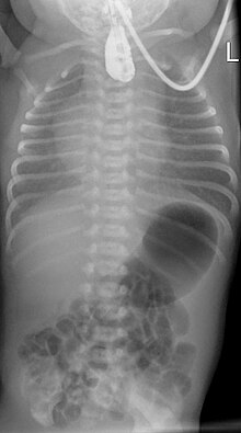
The esophagus is probed for diagnosis . A springy stop is indicative. An X-ray of the chest shows the air filling of the upper blind sac (so-called medallion mark ), and possibly air filling of the intestine as an indication of a lower fistula . In addition, water-soluble contrast media is only given in exceptional cases.
Further malformations must be looked for using ultrasound. For surgical planning, the position of the main artery must be shown in the echocardiogram .
therapy
The operation of a congenital (congenital) esophageal atresia in the newborn was first performed by Cameron Haight in Ann Arbor.
Before an operation, the child's upper body is raised and the secretion is constantly sucked out of the impervious part through a probe.
The surgical correction depends on the distance between the upper part of the esophagus and the lower part. If the distance is small, both parts of the esophagus can be connected to one another with a quick operation. If the distance is large, alternative therapies must be used. Treatment of the esophagus is either extended over several days or weeks until the distance is short enough, or the missing esophagus is replaced by parts of the stomach or intestines that are moved into the chest . If there is a connection to the trachea or lungs , it must be surgically cut and closed to prevent saliva or food from entering the airways.
Course and prognosis
Follow-up treatment is usually necessary over several years. Typical problems are tracheomalacia , gastroesophageal reflux , stenoses at the esophageal suture. Lethality depends on concomitant malformations and the weight at birth. With a birth weight of over 1500 g and without significant heart defects, it is less than 5%.
literature
- Lewis Spitz: Oesophageal atresia . In: Orphanet Journal of Rare Diseases . tape 2 , no. 1 , May 2007, ISSN 1750-1172 , pp. 24 , doi : 10.1186 / 1750-1172-2-24 (English).
- Guidelines
- S2k guideline for short esophageal atresia of the German Society for Pediatric Surgery (DGKCH). In: AWMF online (as of 2012)
Web links
- KEKS patient and self-help organization for children and adults with sick esophagus
- Donauspital: esophageal atresia
Individual evidence
- ↑ Ernst Kern : Seeing - Thinking - Acting of a surgeon in the 20th century. ecomed, Landsberg am Lech 2000. ISBN 3-609-20149-5 , p. 300.
- ↑ M. Yagyu, H. Grid, B. Richter, D. Booss: Esophageal atresia in Bremen, Germany - evaluation of preoperative risk classification in esophageal atresia. In: Journal of pediatric surgery. Volume 35, Number 4, April 2000, pp. 584-587, ISSN 0022-3468 . doi: 10.1053 / jpsu.2000.0350584 . PMID 10770387 .


