shoulder blade
The shoulder blade (in Latin the scapula ) forms the rear part of the bony shoulder girdle in humans and the upper part in animals . In humans and other mammals, it is a flat , triangular bone. In birds it is narrow and saber-shaped. It is used to attach an arm (a front limb in four-legged friends , a wing in birds) and as a muscle origin and attachment . The connection to the chest is purely muscular (so-called synsarcosis , from the Greek syn 'together', sarcos 'meat'). There is an articulated connection to the upper arm bone ( humerus ) and collarbone ( clavicula ) - in birds also to the coracoid . It serves to stabilize the shoulder joint and, thanks to its mobility, adapts to the movements of the upper arm.
Surfaces

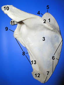
1 Fossa supraspinata, 2 Spina scapulae, 3 Fossa infraspinata, 4 Margo superior, 5 Angulus superior, 6 Margo medialis, 7 Angulus inferior, 8 Margo lateralis, 9 Angulus lateralis, 10 Acromion, 11 Processus coracoidus, 12 origin surface of the teres major muscle, 13 origin of the teres minor muscle
Dorsal facies
The back surface ( Facies dorsalis ) of the shoulder blade (in animals called the side surface - Facies lateralis ) is divided into two pits by the scapulae ( Spina scapulae ): the smaller supraspinous fossa and the larger infraspinate fossa . The origin of the supraspinatus muscle of the same name lies in the supraspinate fossa . The infraspinate fossa located below the spina is largely covered by the infraspinatus muscle , which originates in the two thirds of the pit located in the middle. To the side of this lies the origin of the teres major and the teres minor muscles .
Facies ventralis (costalis)
The ventral surface facing the ribs ( facies ventralis or costalis ) of the scapula shows an extensive concave pit, the subscapular fossa . The two thirds of this pit towards the middle are defined by oblique bone ridges that serve the origin of the subscapularis muscle . The fossa subscapularis is on the lower ( inferior angle (), and upper shoulder blade angle superior Angulus , Syn. Angulus medialis ) by smooth triangular areas from the inner edge ( medial separated). These are called facies serrata in animals and serve to insert the serratus anterior muscle .
Margins
Margo superior
The upper edge ( Margo superior ) is the shortest edge of the shoulder blade in humans. It runs from the superior angle to the base of the coracoid process (Latin: raven beak). In animals, on the other hand, it is significantly longer and points forward, which is why it is called the front edge ( Margo cranialis ). The clear scapular incision ( Incisura scapulae ) is located on the margin of the superior / cranialis and is spanned in humans by a ligament - the ligamentum transversum scapulae superius . The suprascapular nerve runs through this incision . The adjacent part of the superior margin serves as the insertion of the omohyoideus muscle .
Margo lateralis (axillary)
The lateral edge ( Margo lateralis ) is the most massive of all three edges of the shoulder blade. It begins at the lower edge of the joint surface of the shoulder blade ( Cavitas glenoidalis ) and ends at the lower shoulder blade angle ( Angulus inferior ). Directly below the glenoid cavity is a small, rough elevation, the infraglenoid tubercle . This is where the long head ( caput longum ) of the triceps brachii muscle has its origin.
In animals, this edge is directed backwards and is therefore called the rear edge ( Margo caudalis ). This is where the long head of the triceps brachii muscle, teres major and minor muscle and the forearm fascia tensor ( tensor fasciae antebrachii muscle ) arise .
Margo medialis (vertebralis)
The medial margin, which is located towards the center of the body or the spine, is the longest edge of the shoulder blade in humans. It runs from the angulus medialis to the angulus inferior . In animals, it faces the back ( Margo dorsalis ) and is the shortest of the three edges.
Numerous muscles attach to the margo medialis / dorsalis , including the musculus rhomboideus major , the musculus rhomboideus minor and the musculus serratus anterior .
angle
Angulus superior (Angulus medialis)
The upper shoulder blade angle ( Angulus superior , Syn. Angulus medialis ) is created by the meeting of the Margo medialis and Margo superior . It is thin, smooth and rounded and serves as an attachment to some fiber strands of the levator scapulae muscle .
In animals, it is called the anterior shoulder blade angle ( angulus cranialis ) and is located at the meeting of the front and back edges.
Angulus inferior
The lower shoulder blade angle ( Angulus inferior ) is created by the meeting of the Margo medialis and Margo lateralis . It's thick and rough. Its back surface serves as the origin for the teres major muscle and some fiber strands of the latissimus dorsi muscle .
In animals, this angle points backwards and is known as the caudal angle .
Angulus lateralis
The lateral shoulder blade angle ( angulus lateralis ) is the most massive part of the shoulder blade and is also known as the "shoulder head". In animals it points downwards and is called the angulus ventralis . Here lies a flat, cartilage-covered joint socket, the glenoid cavity , which forms the shoulder joint with the head of the humerus .
Prominent structures
Spina scapulae
The shoulder bone ( spina scapulae ) is a compact bone ridge that runs across the back surface of the shoulder blade and divides the shoulder blade into the supraspinous and infraspinous fossa . It begins relatively flat at the medial margin with a smooth triangular surface over which the insertion of the caudal portion of the trapezius muscle slides. On its way to the side, the bone gains height and ends in the acromion, which covers the shoulder joint. Only in horses does the shoulder bone run out flat. In pigs, the shoulder bone has a clear thickening in the middle ( tuber spinae scapulae ).
The trapezius muscle attaches to the upper part of the spine and the deltoideus muscle to the lower part .
Coracoid process
The raven beak process ( processus coracoideus or processus coracoid ) is a strong, hook-shaped bony process of the scapula. It arises from the head of the shoulder blade head and first pulls towards the head and the middle of the body, and then swings to the abdomen and sideways. It tapers off on its way. At the coracoid process of the short head (spring short head ) of the biceps brachii and the muscle coracobrachialis . The pectoralis minor muscle and various ligaments also come into play here.
The coracoid process of mammals is a vestige of the Raven bone ( Os coracoideum ), which is still the strongest bone of the shoulder girdle in birds.
Acromion
The shoulder height or bone corner ( acromion , Greek for ' shoulder bone ', also acromion ) emerges from the shoulder bone and forms the highest point of the shoulder blade in humans. It is not trained in horses and pigs.
The upper surface ( Facies superior ) is roughened and, like the lateral edge of the acromion, serves as the origin for the muscle deltoideus . Here the bone lies directly under the skin and can be palpated as an anatomical reference point . In mammals with a collarbone , on the edge of the acromion in the middle, there is a small oval area that creates the joint connection to it. This joint is known as the ankle joint (articulatio acromioclavicularis) . The acromion serves as the origin of the pars acromialis musculi deltoidei .
In predators (e.g. dog, cat) the acromion is widened to the hamate process . In cats and rabbits, the hamate process also has a hook-shaped appendage pointing backwards ( suprahamatus process ).
Glenoid Cavitas
The scapula joint socket ( Cavitas glenoidalis from Latin cavitas , hollow 'and gleno- , joint socket') is a cartilage-covered, oval joint socket, the vertical diameter of which is larger than the horizontal. Together with the head of the humerus, it forms the shoulder joint . It is surrounded by a cup lip, the glenoid labrum , which deepens the joint socket. The supraglenoid tubercle lies above the socket . This is where the long head of the biceps brachii muscle has its origin.
See also
literature
- Franz-Viktor Salomon et al. (Ed.): Anatomy for veterinary medicine. Enke, Stuttgart 2004, ISBN 3-8304-1007-7 .
Web links
Individual evidence
- ↑ a b Federative Committee on Anatomical Terminology (FCAT) (1998). Terminologia Anatomica . Stuttgart: Thieme.
- ^ Stieve, H. (1949). Nomina Anatomica. Compiled by the Nomenclature Commission elected in 1923, taking into account the suggestions of the members of the Anatomical Society, the Anatomical Society of Great Britain and Ireland, and the American Association of Anatomists, and reviewed by a resolution of the Anatomical Society at the Jena conference in 1935 finally adopted. (4th edition). Jena: Verlag Gustav Fischer.

