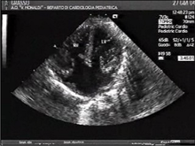
Atrio-ventricular septal defect
| Classification according to ICD-10 | |
|---|---|
| Q21.2 | Defect of the atrial and ventricular septum |
| ICD-10 online (WHO version 2019) | |
The atrio-ventricular septal defect (abbreviation AVSD), also known as AV canal and endocardial cushion defect , is a combined malformation of the heart in the area of the atrium, ventricle and septum, in which there is open connections between the atria and the main chambers. It accounts for about 3% of all congenital heart defects . The frequency is on average 0.19: 1,000 children born alive, with both sexes being affected equally often.
In addition to the atrial septal defect (ASD), this heart defect is often found in connection with Down syndrome (trisomy 21): 43% of all patients with AVSD have Down syndrome (see Borth-Bruns & Eichler: Pädiatrische Kardiologie , 2004, P. 125).
Malformations
The AVSD arises from a hole in the atrial septum ( atrial septal defect ), a hole in the ventricular septum ( ventricular septal defect ) and a malformation of the tricuspid valve (between the right atrium and right ventricle ) and mitral valve (between the left atrium and left ventricle). The aortic valve (between the left ventricle and the aorta ) is further up front than usual. Tricuspid and mitral valves are not actually changed valves, but they have a common valve ring, and this so-called "channel valve" has a variable number of valve leaflets (usually 4 to 7). The excitation conduction system is also relocated due to the defect. This results in the overexcited position type typical for the AVSD in the EKG , mostly in the form of an overexcited left-hand type .
Anatomically, the AVSD distinguishes whether or not there is an effective ventricular septum. This results from whether the level of the connection between left and right is at the level of the heart chambers or just the antechamber. A further distinction is made between AVSD types A, B and C: In addition, the categories complete atrio-ventricular septal defect , with a common canal valve as the only opening between the atria and ventricles and partial or incomplete atrio-ventricular septal defect with separate left and right right openings through a connecting piece of tissue between the pontics at the level of the ventricular septum. If, in addition to a defect in the ostium primum (ASD I), there is a malformation of the inlet valve or an inlet valve, this is referred to as an incomplete AV canal.
Accompanying malformations and / or combinations
can be:
- an underdevelopment ( hypoplasia ) of the right or left ventricle
- a subaortic stenosis (narrowing of the lower part of the aortic valve)
- a coarctation of the aorta
- further ventricular septal defects
- a Fallot tetralogy
Diagnosis
- Echocardiography
- Cardiac catheter examination for surgical planning
- ECG for diagnosing cardiac arrhythmias , overdriven left-hand type (see above)
- X-ray to provide information about the consequences on the pulmonary vasculature
Therapy and prognosis
Most AVSD are operable these days. The circulatory separating operation using the heart-lung machine is accompanied by very good long-term prognoses, depending on the individual characteristics of the AVSD . Repeated operations are the exception. According to current guidelines (ESC 2009), endocarditis prophylaxis is only indicated if a secondary cyanotic heart defect ( Eisenmenger reaction ) has occurred and in the first six months after surgical or interventional treatment of the AVSD using prosthetic material. Regular check-ups are essential.
If left untreated, the AVSD (depending on its severity) can trigger pulmonary hypertension ( high lung pressure ) and finally an Eisenmenger reaction . This causes a reduction in life expectancy . 4 out of 5 children with a complete AV canal do not survive the first two years after birth without treatment (cf. Borth-Bruns & Eichler: Pädiatrische Kardiologie , 2004, p. 131).
The recurrence risk of having another child with AVSD (or another heart defect) in a family is 2.5% if one child is already affected and 10% if two children were born with this heart defect. If the mother has AVSD, the risk of recurrence is statistically 14% (data not confirmed). If the father is affected, 1%. (Both data according to NORA et al. 1991) Since there are special questions for the pregnancy of a woman with a congenital complex heart defect, the absolute number of repetitions should be very low.
literature
- Calabrò R, Limongelli G: Complete atrioventricular canal. Orphanet J Rare Dis. 2006 Apr 5; 1: 8. PMID 16722604 PMC 1459121 (free full text)
- Thomas Borth-Bruhns, Andrea Eichler: Pediatric Cardiology , Springer, Berlin; 1st edition 2004, ISBN 978-3-540-40616-7
Individual evidence
- ↑ Angelika Lindinger, Thomas Paul (Hrsg.): EKG in children and adolescents: EKG basic information, cardiac arrhythmias, congenital heart defects in children, adolescents and adults . 7., completely revised. Ed. Thieme, Stuttgart 2015, ISBN 978-3-13-475807-8 , page 87 .
