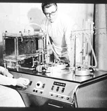
Life-support-machine
The heart-lung machine (HLM) is a medical device that replaces the pumping function of the heart and the lung functions of oxygenation of the blood and carbon dioxide elimination for a limited period of time, thus enabling open heart surgery. The blood leaves the body via a cannula and tube system ( cardiopulmonary bypass ), is enriched with oxygen and pumped back again, this is referred to as an extracorporeal circulation . In addition, a heart-lung machine can quickly cool and warm a patient using a heat exchange (er) . The HLM is not to be confused with the iron lung , which only supports breathing .
The path of the blood usually runs from the vena cava or the right auricle as well as from the heart chambers and opened coronary vessels in the operating area to the HLM and after filtering, oxygenation and carbon dioxide elimination as well as heating and repeated filtering back via the main artery or a femoral artery. In practice, a distinction is made between different types of bypass (total cardiopulmonary bypass, partial bypass, left atriofemoral bypass, femorofemoral bypass, left heart bypass and right heart bypass).
The heart-lung machine is most frequently used in cardiac surgery . In emergency and intensive medicine, smaller, specialized systems are used as so-called extracorporeal membrane oxygenation ( ECMO ).
history

Maximilian von Frey built the first heart-lung machine with his colleague Max von Gruber at the University of Leipzig in 1885 . However, John Heysham Gibbon is considered to be the inventor of the heart-lung machine , whose machine developed in the USA was used in the operation of an atrial septal defect in a 17-year-old patient in 1953. The discovery of heparin by Jay McLean in 1916 is of central importance for the extracorporeal circulation through the heart-lung machine. Heparin prevents blood clotting, which is an elementary requirement for the operation of a heart-lung machine.
Heart-lung machines still use roller pumps to transport blood , the invention of which dates back to 1934.
The discovery of the oxygen enrichment of the blood goes back to an observation in 1944, when it was observed that the blood flowing back to the patient changed color while performing hemodialysis .
In 1926 the Soviet scientist Sergej Brjuchonenko succeeded in creating the first successful extracorporeal circulation on a severed dog's head, whereupon he was the first to predict a future for extracorporeal circulation in cardiac surgery.
After much preparatory work, the American John Gibbon achieved the first extracorporeal circulation on a human on May 6, 1953. He operated on an 18-year-old woman with an atrial septal defect , with the patient connected to the heart-lung machine for 45 minutes. The heart-lung machine was then further developed by Viking Olof Bjork in Sweden and others, among others (cf. Clarence Crafoord and Åke Senning ). In the USA, John Webster Kirklin in particular operated the further development at the Mayo Clinic and used it in 1955 for open heart operations.
With the use of the heart-lung machine, a central problem in cardiac surgery could be solved that had previously made safe operations on the heart impossible: the lack of operating time. In order to make the inside of the heart accessible for surgical interventions, the large cardiac vessels must be temporarily clamped off, which interrupts the oxygen supply to the brain and thus limits the operation time to a few minutes without aids. The mechanical diversion and the oxygenation of the blood played a decisive role in extending this period of time to up to an hour and in operating without haste.
Since the oxygenators used at that time did not achieve the performance of today's devices by far, the blood flow cooling ( hypothermia ) introduced in 1954 with the associated reduction in oxygen consumption was of great importance in order to be able to keep patients alive for a long time with a heart-lung machine .
Around 1955 the construction of an oxygenator succeeded, which enriched blood with oxygen with the help of gas bubbles without the feared danger of air embolism coming into play. In 1956, the type of membrane oxygenator that is still used today was used for the first time . But it would take another 13 years before it was ready for the market.
The first heart operation using a heart-lung machine - created by Manfred Schmidt-Mende and Hans Georg Borst - took place in Germany on February 19, 1958 at the University Hospital in Marburg and was carried out by the eminent cardiac surgeon Rudolf Zenker . A 29-year-old patient with a ventricular septal defect was operated on . With the chronic lack of foreign currency in the German Democratic Republic , Karl-Ludwig Schober developed his own heart-lung machine.
Features and accessories
Pump function, pumps
The heart pumps blood through the blood vessels in a pulsating motion. The pumped volume (cardiac output) is constantly adjusted in order to cope with the often strongly changing loads on the organism. The range of regulation for an adult ranges from approx. 5 l / min at rest to approx. 25 l / min under heavy loads.
Roller pumps are still preferably used today for the extracorporeal circuit . Here, a plastic tube lying in a hemispherical cage is pressed out by two opposing pressure rollers of the centrally rotating pump head. The alternative use of centrifugal pumps is technically more difficult and complex. Finger or axial pumps in occluding operation show a significantly higher hemolysis than roller pumps, which is dependent on the strength and duration of the suction generated during the pumping process. The technical requirements result from the regulation options described above, the connection options to the blood circulation and the safety requirements. The pumps are designed for both continuous and pulsatile operation. The adjustable delivery rates are between 0.01 l / min and 10 l / min. The high precision of the pump head ensures the least possible blood damage (with roller pumps, the hemolysis rate depends on the contact pressure of the pump). An electronic control reliably prevents the uncontrolled speed change of the pump head.
Lung function, oxygenators
The main task of the lungs is the gas exchange of oxygen and carbon dioxide . There are optimal conditions for this in the lungs. The diffusion of oxygen and carbon dioxide takes place over a very large area of up to 200 m², with a thin blood film and a sufficiently long contact time.
The devices available today for saturating blood oxygen ( oxygenators ) can be divided into two classes:
- Bladder oxygenator - gas in direct contact with the blood
- Membrane oxygenator - gas and blood separated
The bubble oxygenator is rarely used in Germany today. But even the membrane oxygenator that is in use today is only imperfectly able to imitate the human lungs. The blood layer is considerably thicker and a diffusion surface of only approx. 2 to 10 m² is available. In addition to gas exchange, today's devices often also take on the function of heat exchangers, so that the blood can be cooled or heated.
Filter function, filter
Microembolism has been a known problem since the heart-lung machine was used . The causes of the microembolism can be fibrin clots , including plastic particles that are rubbed off from tube surfaces or seals or, for example, from B. come from the oxygenator. One tries to counteract this by using blood filters . Another important function of the blood filter is the design-related collection and retention of gas bubbles and the buffy coat .
In addition, a hemofiltration or a modified ultrafiltration can be carried out in order to remove water or urinary substances from the blood in case of renal insufficiency or renal failure.
Dehydration increases the hematocrit and hemoglobin level . In addition, the colloid osmotic pressure increases . This leads to a shift of water from the extracellular space to the intravascular route, which reduces edema (especially pulmonary edema ).
Blood volume depot, reservoir
A so-called cardiotomy reservoir is used as a blood volume depot . In the simplest case it consists of a plastic bag, but often a hard-walled, closed plastic pot with a capacity of over two liters. This makes it possible to withdraw volume that is not required from the patient circuit and return it at a later point in time. In addition to collecting blood, the functions of the cardiotomy reservoir also consist of filtering and defoaming blood from the operating area . Since a blood-air mixture can always be sucked in by sucking blood from the operating area, a defoamer is always required in addition to a filter for tissue components.
Possible complications of extracorporeal circulation
- Blood coagulation disorders (due to thrombocytopenia or heparin-induced thrombocytopenia , insufficient cancellation of the heparin effect , overdose of protamine , coagulation factor deficiency , disseminated intravascular coagulation with consumption coagulopathy )
- Disorders of the water and electrolyte balance ( retention of water; low levels of sodium , potassium , calcium or magnesium )
- Hyperglycaemia (especially with blood sugar levels above 300 mg / dl with the risk of osmotic diuresis )
- Embolism (especially the air embolism with the bladder oxygenator and especially with high oxygen partial pressure values)
- Disorders of the lung function
- Kidney function disorders
- Neurological disorders
Monitoring and documentation
Different parameters are recorded depending on the clinic.
Patient data
- EKG
- Arterial blood pressure
- Central venous pressure
- Temperature rectally / ösophagial
- Renal function / urine output
- Different laboratory parameters
Life-support-machine
- Oxygenator
- Main pump = arterial flow rate = cardiac output = cardiac output
- Mammal
- Cardioplegia system
- Cardiotomy reservoir
- Blood filter
- Arterial / venous oxygen saturation
- Hemoglobin , hematocrit , pH , temperature
- Low-level detector monitors the blood level in the cardiotomy reservoir
- Air bubble detector prevents air from entering the circuit
- Various system pressures
- Arterial / venous blood temperature
- Hose system with connection points
It is now common practice to electronically save the resulting data, which also facilitates subsequent evaluation.
Control devices
Various vital parameters of the patient can be influenced with control devices.
- The oxygen and carbon dioxide transfer in the oxygenator can be controlled with a gas mixer and flow meter.
- The main pump replaces the patient's heart and controls cardiac output.
- Hypo- / hyperthermia devices (Heater-Cooler-Units HCU) can regulate the blood temperature and thus also the body temperature of the patient via the heat exchanger (often in the oxygenator).
Miniaturized extracorporeal circulation (MECC)
By reducing the number of essential components (just the pump and oxygenator), certain disadvantages of conventional heart-lung machines can be reduced and new therapy options can be opened up. The lower surface area coming into contact with the blood reduces the physiological inflammation and coagulation reaction. Furthermore, the complexity of the machine is significantly lower, so that permanent support from cardio technicians is not necessary.
MECCs are sometimes used in routine operations, but above all they offer the possibility of temporary support of the heart and lung function in intensive care patients. The system also resembles an ECMO in terms of the pumps and oxygenators used , but the cannulation is veno-arterial. Blood is thus taken from a vein, oxygenated and introduced into the circulation behind the heart by means of a cannula inserted into the aorta.
When used in intensive care in the are operating theater significant disadvantages of the lack of reservoirs (thus stolen the surgical site blood no longer reperfusion) and the lack of additional suction pumps (Vent) is not important.
In principle, the system can be implanted for any type of circulatory failure. Of course, this is only sensible and ethically justifiable if the underlying disease is potentially reversible. The main indications are cardiogenic shock, postoperative pump failure, myocarditis and a bridge-to-decision or a bridge-to-transplantation to bridge the gap until further therapy ( artificial heart implantation or heart transplantation ) .
The size of the devices has now shrunk to such an extent that they can be transported using conventional air and ground-based intensive care vehicles. The extent to which this technology will spread beyond specialized centers is, however, at least questionable due to the complex underlying diseases and the resulting intensive therapy, especially since peripheral cannulation is considered to be technically demanding and the most common source of complications.
Therefore, some centers offer ECMO and MECC support for peripheral hospitals, whereby a team from cardio technology / heart surgery and anesthesia is brought to the peripheral hospital (mostly by air). On site, the patient can be connected to the heart-lung machine and transferred to a center. Often this is the only way to transport patients with unstable circulatory function.
user
In the early days it was the job of a doctor to operate the heart-lung machine. Today this is done by the cardio technician. Initially, you learned the profession part-time. There were z. B. Surgical nurses or medical technicians trained.
With the increasing area of responsibility and increasing complexity of the tasks, however, the need for targeted training was recognized. This has been mainly taken over by the Akademie für Kardiotechnik in Berlin since 1988 , which has offered a practice-oriented bachelor's degree since 2008 and is the only institute in Germany to have state recognition.
In 1994, the first major cardiac engineering major was established at the Aachen University of Applied Sciences (Jülich Department), which was later followed by the "Medical Engineering" course at Furtwangen University .
As a professional association, the Deutsche Gesellschaft für Kardiotechnik has taken on the representation of interests in Germany , and in Europe the EBCP (European Board of Cardiovascular Perfusion).
literature
- Susanne Hahn: Heart-Lung Machine (HLM). In: Werner E. Gerabek , Bernhard D. Haage, Gundolf Keil , Wolfgang Wegner (eds.): Enzyklopädie Medizingeschichte. De Gruyter, Berlin / New York 2005, ISBN 3-11-015714-4 , p. 584.
- Reinhard Larsen: Anesthesia and intensive medicine in cardiac, thoracic and vascular surgery. (1st edition 1986) 5th edition. Springer, Berlin / Heidelberg / New York a. a. 1999, ISBN 3-540-65024-5 , pp. 79-120 ( cardiopulmonary bypass ) and 139-165 ( practical procedure for operations with the heart-lung machine ).
- Wolfgang Eichler, Anja Voss: Operative intensive care medicine. In: Jörg Braun, Roland Preuss (Ed.): Clinic Guide Intensive Care Medicine. 9th edition. Elsevier, Munich 2016, ISBN 978-3-437-23763-8 , pp. 619–672, here: pp. 654–660: Interventions with a heart-lung machine (HLM) .
Web links
- herz-lungen-maschine.de
- Website of the Academy for Cardiac Technology at the German Heart Center Berlin
- Website of the German Cardiac Society
- European Board of Cardiovascular Perfusion website
- For the first time in air use: The world's smallest portable heart-lung machine at innovations-report.de
Individual evidence
- ↑ Reinhard Larsen: Anesthesia and intensive medicine in cardiac, thoracic and vascular surgery. 1999, p. 80 f. and 117-120.
- ^ J. Willis Hurst, W. Bruce Fye, Heinz-Gerd Zimmer: The heart-lung machine was invented twice — the first time by Max von Frey . In: Clinical Cardiology , Vol. 26, September 2003, pp. 443-445, doi: 10.1002 / clc.4960260914
- ↑ Susanne Hahn: Heart-Lung Machine (HLM). 2005, p. 584.
- ^ Benjamin Prinz: Operating on the Bloodless Heart: A History of Surgical Time Between Crafts, Machines and Organisms, 1900-1950 . In: NTM Journal for the History of Science, Technology and Medicine . tape 26 , no. 3 , 2018, p. 237-266 , doi : 10.1007 / s00048-018-0195-x .
- ^ Hans-Jürgen Peiper : The Zenker School. (Address on the occasion of the ceremony for the 68th birthday of Prof. Dr. med. Horst Hamelmann on May 26, 1992 in Würzburg) In: Würzburger medical history reports 11, 1993, pp. 371–387, here: pp. 379 f.
- ↑ Reinhard Larsen: Anesthesia and intensive medicine in cardiac, thoracic and vascular surgery. 1999, p. 82 f. and 107.
- ↑ Reinhard Larsen: Anesthesia and intensive medicine in cardiac, thoracic and vascular surgery. 1999, pp. 110-113.
- ↑ Reinhard Larsen: Anesthesia and intensive medicine in cardiac, thoracic and vascular surgery. 1999, p. 113.
- ↑ Reinhard Larsen: Anesthesia and intensive medicine in cardiac, thoracic and vascular surgery. 1999, p. 114.
- ↑ Reinhard Larsen: Anesthesia and intensive medicine in cardiac, thoracic and vascular surgery. 1999, p. 114.
- ↑ Reinhard Larsen: Anesthesia and intensive medicine in cardiac, thoracic and vascular surgery. 1999, p. 114 f.
- ↑ Reinhard Larsen: Anesthesia and intensive medicine in cardiac, thoracic and vascular surgery. 1999, p. 115.
- ↑ Reinhard Larsen: Anesthesia and intensive medicine in cardiac, thoracic and vascular surgery. 1999, p. 115 f.

