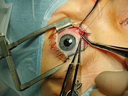
Eye surgery
The ophthalmology knows many surgical methods for the treatment of organic or functional disorders, and for correction of movement disorders and optical refractive errors of the eye .
Cataract operations
During cataract operations , the clouded eye lens is removed and usually replaced with an artificial lens . In general, the surgical removal of the lens of the eye is also known as a lensectomy . The cataract operation is one of the most frequently performed surgical interventions. The following methods are used:
The intracapsular cataract extraction (ICCE) is only used in exceptional cases. The lens including its capsule is removed. An artificial lens is not used (the patient is given cataract glasses ) or positioned on the iris (iris-supported), in the chamber angle (as an anterior chamber lens) or sewn to the dermis as a so-called sclera-fixed lens.
With extracapsular cataract extraction (ECCE), the lens capsule is left in place and the artificial lens is inserted in the capsular bag or, more rarely, in front of the capsule (sulcus-fixed lens). A distinction is made between the seldom used classic method of manual extraction of the nucleus (delivery) and so-called phacoemulsification . The phacoemulsification is the most widely used in Germany technology. The lens is smashed with a tube vibrating at an ultrasonic frequency and suctioned off. The cut on the cornea is therefore much smaller than at delivery and is only approx. 2 mm.
The YAG laser capsulotomy enables the treatment of the "secondary cataract", which can occur in approx. 20–30% of patients after cataract surgery. The lens capsule, which is cloudy due to fibrosis, is opened and the cloudiness is removed.
In recent years, femtosecond laser cataract surgery has developed into a gentle and precise laser-supported surgical procedure for the treatment of cataracts.
Glaucoma operations
During surgery for glaucoma (glaucoma), depending on the type of disease, the drainage of the aqueous humor is improved and the intraocular pressure is thereby reduced. The following methods are used, among others:
- Goniotomy or angulocision: incision of the tissue in the drainage area of the aqueous humor to improve the drainage, e.g. B. in congenital forms of glaucoma (developed and published in 1953 by the American ophthalmologist Otto Barkan from Vienna).
-
Iridotomy : creation of a flow opening in the iris to improve the aqueous humor circulation in narrow-angle glaucoma .
- Laser iridotomy without opening the eyeball
- surgical iridectomy , through an incision on the edge of the cornea, or during other eye surgery.
-
Trabeculotomy , incision of the trabecular structure to improve the drainage of the aqueous humor.
- Excimer laser trabeculotomy from interno
- Trabeculectomy and other filtering interventions, creating an artificial opening in the sclera , which is then loosely closed and covered with conjunctiva.
-
Microinvasive glaucoma surgery (MIGS): OP techniques that have become increasingly popular in recent years and that have the great advantage of being able to be carried out on an outpatient basis, as the risks are significantly more manageable. In terms of pressure reduction, MIGS are not as successful as classic trabeculectomy. Drip therapy usually has to be carried out after the operation.
- XEN stent (drainage under the conjunctiva as in a trabeculectomy with the formation of a filter cushion)
- iStent (trabecular bypass: micro-implant placed in the trabecular network)
- Cypass implant (aqueous humor is directed to the choroid: "uveoscleral drainage") - Cypass was withdrawn from the market in September 2018 due to the long-term risk of possible corneal damage.
- Cyclodestructive interventions, destruction of part of the ciliary body that produces the aqueous humor.
- Cyclophotocoagulation with the laser (CPK)
- Cyclocryocoagulation, with cold (cryo)
-
Trabeculoplasty , stretching of the meshwork in the drainage area of the aqueous humor through targeted placement of scars in the adjacent tissue. Not in all cases long-lasting pressure reduction
- Argon laser trabeculoplasty (ALT)
- selective laser trabeculoplasty (SLT)
Corneal surgery
- penetrating keratoplasty (PK) - corneal transplantation is used to replace a diseased cornea with a donor cornea.
- Lamellar Keratoplasty - Replacing only one layer (lamella) of the cornea with donor tissue
- Keratoprosthesis (partly experimental) - Insertion of non-corneal (not belonging to the cornea) material (artificial, tooth or similar) as a corneal replacement.
- Phototherapeutic keratectomy (PTK) - targeted vaporization of the clouded corneal tissue using a laser.
- Pterygium removal, excision of the pre-growing tissue (with / without conjunctival transplantation) or antimetabolites
- Keratography (corneal tattoo) - cosmetic reconstruction procedure for a number of clinical pictures
Vitreous and Retinal Surgery
-
Vitrectomy , removal of the vitreous as much as possible
- Pars plana vitrectomy (PPV), through access through the zone between the retina and the ciliary body
- with or without peeling epiretinal membranes
- with or without vitreous replacement by gases or special liquids (e.g. silicone oil)
- with removal of the vitreous boundary membrane (ILM peeling)
- Open-sky vitrectomy, through access via the previously removed cornea
- Pars plana vitrectomy (PPV), through access through the zone between the retina and the ciliary body
- Retinal rotation in macular degeneration
- Targeted placement of retinal scars to improve attachment or to reduce the need for oxygen and nutrients.
- Photo or laser coagulation
- Cryocoagulation, with cold
- Denting operation for detachment of the retina ( hump surgery), denting of the sclera from the outside, e.g. B. to close a retinal hole
- scleral seal, mostly spongy plastic
- Pneumatic retinopexy, entry of air into the vitreous space to (additionally) close the retinal hole from the inside.
- Cerclage with or without a wedge, plastic tape that is placed around the eyeball like a belt.
- scleral seal, mostly spongy plastic
- Sclerochoroidectomy (block excision) for tumors of the choroid
Refractive surgery
The refractive surgery is used for the correction of refractive errors ( ametropia ) of the eye (the methods for details see refractive surgery ). The following methods are known:
- Keratomileusis
- automated lamellar keratoplasty (ALK)
- laser assisted in-situ keratomileusis ( LASIK )
- laser assisted sub-epithelial keratomileusis ( LASEK ), also known as Epi-LASIK
- photorefractive keratectomy ( PRK )
- Laser Thermo Keratoplasty (LTK)
- conductive keratoplasty (CK)
- Limbal Relaxing Incision (LRI)
- anterior ciliary sclerotomy (ACS)
- astigmatic keratotomy (AK), or arcuate keratotomy or transverse keratotomy
- radial keratotomy ( RK )
- hexagonal keratotomy (HK)
- Epikeratophakia
- corneal ring segments ( Intacs )
- implantable artificial lenses
- as “piggyback lenses” between the iris and the lens
- as iris clip lenses on the front of the iris
- scleral expansion ligaments
- KAMRA implant
Eyelid surgery
- Ptosis operation by folding the levator palpebrae superioris muscle or levator suspension by suspending the lid near the browbud
- surgical removal of chalazion and tumors
- Sliding sculpture (Tenzelplastik)
- tarsomarginal transplants (according to Huebner)
- free skin grafts
- Upper and lower eyelid extension
- Eyelid cleft reductions
- Tarsoraphy (sewing of the upper and lower eyelid)
- Blepharoplasty (plastic skin incision)
- Laser treatment to remove xanthelasma
Tear duct operations
- endoscopic lacrimal duct surgery
- Lacrimal duct surgery according to Toti
- Permanent probe according to Remky
Eye muscle surgery
Operations on the eye muscles are performed to correct strabismus ( strabismus ), eye tremors ( nystagmus ) and ocular constrained head postures . The surgical correction of the squint is also known as a squint operation . It can be necessary at any age and can be considered if the extent of a squint angle does not allow the development or restoration of binocular vision or makes central fixation impossible. Cosmetic considerations also play a role. Different procedures influence the functioning of one or more eye muscles. The following principles form the basis for this:
- Change in muscle strength
- Change in excursion ability
- Change of the rolling distance
- Change in the position of the eyeball
- Change in the direction of muscle pull
As a rule, the aim of such operations is primarily a functional improvement and only in a second respect a cosmetic one .
Oculoplasty
- Blepharoplasty, numerous surgical procedures to correct the eyelid skin or eyelid position.
- Removing the eyeball
- Enucleation - removing the eyeball and leaving the eye muscles and the rest of the orbital contents with or without inserting a guide seal for an artificial eye that will later be placed on the outside .
- Evisceration - removal of the contents of the eyeball, leaving the sclera intact. To reduce pain in a blind eye.
- Exenteration - removal of the eye and orbital contents, including extraocular muscles, fat and connective tissue; usually with malignant tumors .
Part of the specialist training
In order to be admitted to the ophthalmological specialist examination, the doctors must prove that they have fulfilled an examination, treatment and operation catalog. This is listed in the documentation of the advanced training according to the (sample) advanced training regulations (MWBO) .
literature
- Brigitte Lengersdorf, Detlef Rose: Ophthalmology (ophthalmology) . In: Margret Liehn, Brigitte Lengersdorf, Lutz Steinmüller, Rüdiger Döhler (eds.): OP manual. Basics, instruments, surgical procedure . 6th, updated and expanded edition. Springer, Berlin / Heidelberg / New York 2016, ISBN 978-3-662-49280-2 , pp. 705–718.
- O. Haab: Atlas and outline of the doctrine of eye operations . Bremen University Press, reprint of the original edition from 1904, ISBN 3-95562-559-1
Web links
- Federal Association of German Ophthalmic Surgeons e. V.
- Sk2 guideline for visual perception disorders (PDF) of the AWMF , status 04-2017; accessed on January 26, 2018
- Patient information on laser-assisted cataract surgery
Individual evidence
- ↑ Guideline No. 19 a from BVA and DOG - Cataract surgery (cataracts) in adulthood
- ↑ Information document ( Memento from June 25, 2012 in the Internet Archive ) (PDF; 401 kB) Federal Association of Ophthalmologists in Germany BVA
- ↑ Guideline No. 19 b by BVA and DOG - Nd: YAG laser capsulotomy of the cataract
- ↑ Burkhard Dick, Ronald D. Gerste, Tim Schultz: Femtosecond Laser in Ophthalmology. Thieme, New York 2018, ISBN 978-1-62623-236-5
- ↑ Wolfgang Leydhecker: Advances in modern ophthalmology. In: Würzburg medical history reports. Volume 3, 1985, pp. 185-210, here: p. 195.
- ↑ Alcon announces voluntary global market withdrawal of CyPass Micro-Stent for surgical glaucoma | Alcon.com. Retrieved November 18, 2018 .
- ↑ Herbert Kaufmann (Ed.): Strabismus . 5th completely revised edition with Heimo Steffen. Georg Thieme Verlag, 2020, ISBN 978-3-13-241330-6 .
- ↑ Guideline No. 26 c from BVA and DOG - eye muscle surgery due to non-paretic strabismus and nystagmus-related head posture
- ↑ Guideline No. 27 a from BVA and DOG - eye muscle surgery due to paretic strabismus
- ↑ German Medical Association Documentation of the advanced training according to the (sample) advanced training regulation (MWBO) on the specialist training in ophthalmology , version of June 26, 2010 and February 18, 2011 (PDF)


