Excitation conduction system
|
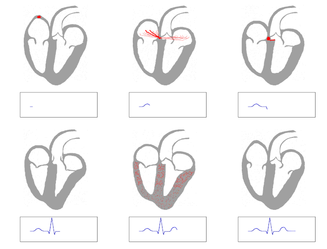
Excitation conduction system (schematic, in humans) 1 sinus node - 2 AV nodes Important structures are |
The conduction system of the heart, passes the electrical signals which regulate the pumping action of the heart. The basic rhythm of these impulses is generated by the stimulation system. Both systems - the excitation and conduction system - of the heart do not consist of nerve cells , but of specialized heart muscle cells .
Arousal education system
The sinus node serves as the primary pulse generator (pacemaker) of the heart . The sinus node generates electrical impulses. Due to the position of the sinus node in the wall of the right atrium at the junction of the superior vena cava , the electrical excitation and thus the contraction of the muscle cells comes from the right atrium. The sinus node produces around 60 to 80 excitations per minute.
The secondary pacemaker of the heart is the atrioventricular node or AV node for short. In the event of a failure of the sinus node, the AV node can take over the pulse generation as a secondary pacemaker. The AV node itself can "generate" 40 to 50 excitations per minute. However, since this frequency is exceeded by that of the sinus node in the healthy heart, its pacemaker activity is not used. In a so-called AV block , in which the conduction through the AV node is partially or completely disturbed, the AV node acts as a pacemaker for the heart.
In principle, every heart muscle cell has the ability to generate excitation ( automatism ).
Excitation conduction system
The excitations are passed on by the His-Purkinje system . First they get from the AV node to the bundle of His (according to Wilhelm His ). The bundle of His also has its own rhythm and can initiate 20 to 30 excitations per minute. As a tertiary pacemaker of the heart , the bundle of His can thus assume a backup function for the AV node.
The common trunk of the bundle of His ( Truncus fasciculi atrioventricularis ) is divided into three “branches”: Two left and one right tawara limb (after Sunao Tawara ). If the conduction of excitation is delayed or interrupted in one of the thighs, one speaks of a bundle branch block ( right or left bundle branch block ). At the apex of the heart, the thighs are further divided into Purkinje fibers or Myofibrae conducens Purkinjiensis (after Jan Evangelista Purkinje ), which represent the last conduction routes of the excitation conduction system and come into contact with the cardiac muscle fibers of the working muscles. The abundant glycogen in the cells of the Purkinje fibers is worth mentioning. In addition, a few muscle fibrils can be detected under the cell membrane .
Definition of terms: stimulus and arousal
A stimulus (e.g. heat, pressure, pain, etc.) is an external effect that is absorbed, for example, in the skin by sensory cells (receptors). A stimulus causes electrical impulses to develop in the downstream nerve cells, which are known as excitation. However, no stimulus is necessary for the generation of excitation in the heart and the conduction of excitation through the fibers of the excitation conduction system. Only the electrical impulses (not the stimulus itself) are passed on by the fibers.

literature
- HG Borst, W. Klinner, A. Senning (Ed.): Heart and vessels close to the heart. Springer Verlag, Berlin / Heidelberg 1978, ISBN 978-3-662-07752-8 .
- W. Hort (Ed.): Pathology of the endocardium, the coronary arteries and the myocardium. Springer Verlag, Berlin / Heidelberg 2000, ISBN 978-3-642-62944-0 .
- Richard Nickel, August Schummer, Eugen Seiferle (Ed.): Circulatory system, skin and skin organs. 3rd edition, Parey Verlag, Stuttgart 1996, ISBN 3-8304-4164-9 .
See also
Web links
- Conduction system of the heart. DocCheck Flexikon
- Physiology of the heart. (PDF, 259K) University of Münster (with a description of the classic experiments on the "virtual" rat heart).
- The conduction system of the heart. (PDF, 938K) Samaritan Association of the Canton of Solothurn(presentation of a training course).
- Ina Michel-Behnke (Department of Pediatric Cardiology, University of Giessen): Living with a chamber: the univentricular heart. (PDF, 48K) Herzkind e. V.
Individual evidence
- ^ Walter cherry : Jan Evangelista Purkyně 1787–1868. A contribution to the 200th anniversary of his birthday. Akademie-Verlag, Berlin 1989 (= session reports of the Academy of Sciences of the GDR. Year 1988, No. 5 / N), ISBN 3-055-00520-1 , p. 30.

