canine

The canine ( latin the canine , plural Denies canini , often Canini ) is the cone-shaped tooth in the teeth of mammals (including humans) behind the front teeth (incisors) and before the Vorbackenzähnen (premolars). In the upper jaw , the canine is the foremost tooth in the upper jawbone (maxillary) after the intermaxillary bone (premaxillary ).
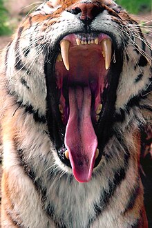
The term "canine" refers to the clear kink of the dental arch at this position. Another name is "dog tooth" ( adjective caninus "doggy", "from the dog"). In land carnivores , the canine is enlarged to a fang due to its function in the acquisition of prey .
human
designation
The translation of the Latin dens caninus as "dog tooth" occurs in a number of other languages, analogous to German. In the case of inflammation of an upper canine, the inflammation in the face can manifest itself with swelling, reddening and tenderness just below the eye, as the tip of the upper canine reaches almost to the orbit (bony eye socket). Hence the old name eye tooth . In some other languages, too, the tooth is called accordingly (for example Icelandic Augntönn or English eye tooth ).
Evolution of the human canine
In the evolution of mammals , the canines have generally changed little - they are always single-pointed and single-rooted , both in the upper and lower jaw. The shape of the human canines differs from that of the other primates . In many primates, including humans, the canine tooth is enlarged in males; this sexual dimorphism is particularly pronounced and the like. a. in the great apes (Hominidae). The upper canines are much more elongated than the lower ones. A function in the threatening gesture is assumed to be the main cause of this development .
In the human dentition, the upper canines have the longest roots. Since the tip part of these teeth is considerably longer than the root part of all primates except humans, a considerable shortening of the crown is assumed in the course of hominization (" becoming human "). The upper canines of the non-human primates show a clear tendency to match the shape of the following molar teeth (premolarization), while the lower canines morphologically approach the incisors (incisivation). In humans, all canines show an incision, which is more pronounced in the lower jaw.
dentition
Humans have one canine tooth per half of the jaw in the lower and upper jaw (a total of four). The canine is in the third position (counting from the first incisor) and is the largest tooth in the anterior region. The canines are the cornerstones at the transition from the front teeth to the posterior teeth.
The canine is already created in the deciduous dentition, the tooth eruption occurs at around 1.5 years of age.
The eruption of the permanent canines occurs at around 11 years of age (± 1 year). Usually the lower canines erupt before the upper canines. The exact breakthrough times vary depending on the release:
- upper permanent canines: boys 11.8 ± 1.3 years; Girls 11.2 ± 1.2 years
- lower permanent canines: boys 11.1 ± 1.6 years; Girls 10.2 ± 1.3 years
Most of the time, after the incisors in the upper jaw, the anterior premolars break through before the canines break through. It is the other way around in the lower jaw. Hence the frequent lack of space for the upper canines (see below).
root
The canine has a root that contains a canal. The root is slightly flattened in the mesial-distal direction (from the center of the jaw to the rear, i.e. away from the center). In the upper canines, the mesial root surface is wider and flatter than the distal root surface.
The upper canines have a distinct root feature with an apical (at the tip) curvature distally. Both of the lower canines are missing.
The roots of the lower canines are shorter than those of the upper. The length relation between crown length and root length is shifted in favor of the crown length in the lower canines.
Dental crown
Instead of a chewing surface, the canine only has a cusp tip (canine tip) with two short cutting edges.
While the outer (vestibular) surfaces of the incisors are relatively flat apart from a slight curve, the vestibular surfaces of the canines are divided into two parts, a mesial (front) and distal (rear) half. Both halves form an angle of approx. 20 ° to each other. They are separated by a vertical central ridge. The dental arch kinks at this point.
In addition, the canine, like all teeth, has a slightly spherical shape. It has a slight convexity (curvature) from the incisal edge to the tooth neck.
The more or less sharp point on the incisal edge (canine tip) is not exactly in the middle of the incisal edge, but is shifted a little mesially. The mesial incisal edge is steeper and shorter than the flatter and longer distal incisal edge.
On the back, the canine crown has two strongly developed marginal ridges and a central ridge that meet in a pronounced cusp (tuberculum) towards the neck of the tooth.
The approximal surfaces (contact surfaces with the neighboring tooth) are triangular.
The lower canines are smaller than the upper canines. In the lower canines, the crown axis is somewhat “bent” lingually (on the tongue side) compared to the root axis (“crown alignment”). This crown alignment can also be found in all premolars and molars in the lower jaw. The vestibular (lip / cheek side) surface has an incline of 25 ° compared to the root surface. The mesial contact surfaces are steeper, with the enamel-cement boundary 3 mm higher mesially than distally.
Diseases

In addition to the usual dental diseases such as tooth decay , pulpitis and apical ostitis , the upper canine is very often retained and displaced.
The reason for this is the relatively late breakthrough time of eleven years. At this age, the bones of some children are already quite solid. In addition, the permanent neighboring teeth (second incisor and first premolar) are already there before the canine and, if there is a pronounced lack of space, they can take up all the space for the canine that erupts late. The canine may also break through further vestibularly due to lack of space - outside the row of teeth. It then protrudes like a tiger's tooth from the vestibular wall of the alveolar process .
The impacted canine tooth is relatively often transversely impacted in the maxillary bone.
Another cause of retention is that the canine is relatively high up in the jawbone during its formation phase and has to cover a very long way to eruption.
After the wisdom teeth, the canines are the second most frequently affected teeth that are retained and dislocated. A persistent milk canine in the upper jaw indicates a retained canine. A failure of the canines is not known or extremely rare. In contrast to this, a non-abutment is often found in wisdom teeth (approx. 50%) and occasionally (approx. 1%, clustered in families) in the neighboring second incisors of the upper jaw.
The retained and displaced upper canines are usually surgically removed in adults for orthodontic indications (usually with a palatal surgical approach). In adolescents and if the vestibular retention position is favorable, the tooth crown is surgically exposed and, after the wound has healed, it is adjusted with the help of a glued-on bracket and a fixed or removable orthodontic device . Both multiband appliances and simpler orthodontic appliances are used here. In some cases, sufficient space must be created for this by stretching the upper jaw ( KFO ). The tooth is looped onto the bracket and mostly successfully integrated into the row of teeth over the course of a few months or years.
Lower canines are less often retained and dislocated than upper canines.
Malformation
Typical malformations occur in connection with cleft lip and palate . The cleft lip typically runs between the 2nd and 3rd tooth in the upper jaw - that is, between the second incisor and canine. In the case of abortive forms of cleft lip, which clinically do not express themselves as a cleft lip, there may be fusions, partial fusions or adhesions of the 2nd and 3rd tooth or these teeth may be affected individually or there may be an additional tooth between the 2nd and 3rd teeth. and 3rd tooth occur. This surplus tooth usually has a narrow cone shape or is crippled.
Occasionally, adhesions between the 2nd and 3rd tooth can also be observed in milk teeth. With milk teeth, these adhesions also occur in the lower jaw, which calls into question the suspected connection with cleft lip.
Canine guidance
At rest with closed rows of teeth, the upper and lower molars of one side touch. With lateral chewing movements, there is inevitably a gap between the upper and lower molar chewing surfaces, since the upper and lower canines first collide and, as it were, force the rows of teeth apart as the first "obstacle". This so-called canine guidance is part of the complicated interplay between the chewing surfaces, jaw joints and masseter muscles that gnathology deals with. Often this leadership property exists together with the premolars (premolar guidance ). The canines are anatomically predestined for this due to a larger crown-root length ratio. As a result, the canines absorb the lateral chewing forces that would otherwise act pathologically on the molars. The latter can lead to tooth loosening.
When manufacturing fixed dentures ( crowns , bridges ), the canine guidance must be restored as far as possible.
In the manufacture of full dentures, on the other hand, no canine guides may be created, as the point-like contact between the upper and lower denture canine teeth would tilt the full denture. For the benefit of a stable chewing function, guidance is created when the lower jaw moves sideways through all molars (premolars plus molars) on both sides.
aesthetics
The presence of the upper canines is important for the aesthetically natural appearance of the front teeth. If space has to be created for crowded teeth as part of an orthodontic treatment, the first premolars are usually extracted. This also takes place if a displaced canine needs to be adjusted.
The opposite problem arises when the upper lateral incisors are not applied and the canines follow next to the first incisors. For aesthetic reasons, in these cases the canine is optically changed to a lateral incisor. This is done by grinding the cusp tip of the canine tooth and building a cutting edge on it using composite materials. Alternatively, the canine can be reshaped using a veneer .
In contrast to European ideals of beauty, relocated upper canines are considered cute in Japan, especially among girls, and there are called Yaeba ( Japanese 八 重 歯 , dt. "Multiple teeth"). This phenomenon also occurs relatively often there, due to the smaller jaw and because the teeth are seldom straightened.
Toothing (antagonists)
The upper canines are in contact with the lower canines and the first premolars of the lower jaw behind them.
The lower canines are in contact with the upper second incisors and the upper canines.
Other mammals

Most mammals also have two canines in the upper jaw and two in the lower jaw . In horses , usually only stallions have canine teeth, which are referred to here as hook teeth . Rabbits and rodents do not have canine teeth. In many ruminants , they are absent in the upper jaw. In walrus and hippopotamus , the canines form the tusks , in pigs the weapon and in predators the fangs . The space between the upper canine tooth and the first incisor is called the diastema , in primates it is also known as the “ape gap”. The canines of monkeys - including the great apes - are in relation to the incisors significantly greater than in anatomically modern humans ( Homo sapiens ), so the "monkey gap" is required. A fossil human skull can be distinguished from an ape skull in that this ape gap closes in the prehistoric and early humans and the incisors form a continuous dental arch with the not so large human canines. In humans, a diastema occurs particularly between the two incisors and is called diastema mediale .
See also
Web links
Individual evidence
- ^ Ulrich Lehmann: Paleontological Dictionary . 4th edition. Ferdinand Enke Verlag, Stuttgart 1996, p. 39 .
- ↑ See, for example, Max Höfler: German Name of Disease Book. Munich 1899, p. 840.
- ↑ a b Winfried Henke , Hartmut Rothe : Paläoanthropologie . Springer-Verlag, Berlin / Heidelberg / New York 1994, p. 127-129 .
- ↑ Albert Mehl, Karl-Heinz Kunzelmann, Veneers ( Memento of the original from July 22, 2013 in the Internet Archive ) Info: The archive link was inserted automatically and has not yet been checked. Please check the original and archive link according to the instructions and then remove this notice. , BLZK, ZBay 1-2 / 2001
- ↑ Austin Considine: A Little Imperfection for That Smile? In: The New York Times . October 23, 2011, p. ST6 ( online ).
- ^ Emil Kuhn-Schnyder, Hans Rieber: Paläozoologie. Thieme Verlag, 1984, ISBN 3-13-653301-1 , pp. 280-286.
- ↑ Donald Johanson , Edgar Blake: Lucy and her children. Spektrum Verlag, 2000, ISBN 3-8274-1049-5 .
- ↑ Wolfgang Schad : Gestalt motifs of fossil human forms. In: Goethean Natural Science, Volume 4 Anthropology. Stuttgart 1985, pp. 111-112.


