Single molecule experiment

A single molecule experiment ( in English: single-molecule experiment ) is an experiment looking at the properties of single molecules. Single molecule experiments are in contrast to measurements of a totality or accumulation of molecules, in which the behavior of individual molecules cannot be distinguished and only average values are measured. While many experiments in biology, chemistry, or physics are not sensitive enough to observe single molecules, there are single molecule fluorescence techniques that have generated great excitement as they reveal new details that were not visible with previous measurement techniques. Indeed, many techniques have been developed to detect single molecules since the 1990s.
The first single molecule experiments were patch clamp experiments carried out in the 1970s, but these are limited to measurements on ion channels . Today, single-molecule techniques are used, which can measure the movement of myosin on actin filaments in muscle tissue, and investigate the spectroscopic details of the environment in solutions. Biological polymers have been studied with atomic force microscopes . Individual molecules and their elastic deformation are measured microscopically, usually in the polymer.
history
Single molecule experiments have been carried out for decades at low pressure, but only since 1989 by William E. Moerner and Lothar Kador also in condensed phases. A year later, Michel Orrit and Jacky Bernard were able to detect the absorption of single molecules based on their fluorescence. There are many techniques for observing single molecules, such as mass spectrometry, where single ions are detected. In addition, there is single molecule detection in the field of ion channels with the development of the patch clamp technique by Erwin Neher and Bert Sakmann . The idea of using conductivity measurements to detect individual molecules had reached its limits. Fluorescence measurements, on the other hand, are suitable for observing a single molecule thanks to the sensitivity of optical detectors that are able to count individual photons . The observation of individual molecules with spectroscopic methods requires an isolated environment and the emission of photons with photochemical excitation , which includes equipment (photomultiplier or extended photodiodes) with which one is able to record photoemissions with high sensitivity and high time resolution.
Recently, single-molecule fluorescence has been the subject of interest in biological imaging, since the labeling of biomolecules such as proteins and nucleotides to explore enzymatic functions cannot be done in an overall structure because of the subtle time-dependent movements involved in catalysis and structural reorganization. The most widely studied protein is the myosin / actin enzyme, found in muscle fibers. Using the single molecule technique, many individual reaction steps were observed and many of these proteins were characterized.
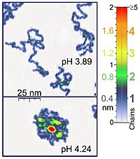
Atomic force microscopes (atomic force microscope (AFM)) are also available for single molecule experiments of biological significance because they work in the same range as most biological polymers . In addition, AFMs can be used for synthetic polymers, and they can map polymer chains in 3D. the AFM is fine enough to observe absorbed polyelectrolytes (e.g. poly-2-vinylpyridine) in solution. If two chains overlap, one has twice the thickness of a single chain. If the scan parameters are set correctly, the conformation of the chains is retained for hours and allows the experiments to be carried out in solution under the appropriate reaction conditions. If you check the force field around the sample, you get high-resolution images. Optical tweezers have also been used to measure DNA-protein interactions.
Experiments
concept
Single molecule fluorescence spectroscopy uses the fluorescence of a molecule to obtain information about the environment, structure and position. Technology opens up the possibility of obtaining information that cannot be obtained by averaging an overall structure. In most experiments with single molecules the results are averaged.
Single channel recording
As in the case of single-molecule fluorescence spectroscopy, this technique, known as single-channel recording, is used to record specific kinetic information - the ion channel function - that cannot be read from the overall structure, e.g. cell recordings. Ion channels alternate between conducting and non-conducting states, depending on the conformation. Therefore, the functional state of an ion channel can be measured. Single molecule experiments are carried out systematically under different reaction conditions, so that the different kinetic states of the ion channel can be observed.
Biomolecule marking
Individual fluorophores can be chemically linked to biomolecules, such as proteins and DNA , and the movement of the individual molecules can be recorded with the aid of the fluorescent marker. Molecular movements can be detected based on the change in emission intensity or radiation duration, which indicates changes in the environment. For example, single molecule labeling has provided a lot of information about kinesin proteins in microtubules of muscle cells.
Single Molecule Fluorescence Resonance Energy Transfer ( FRET )
In single molecule fluorescence resonance energy transfer (FRET), the molecule is marked in two places. A laser beam is radiated onto the first sample for excitation. As soon as this sample relaxes and emits a photon, it can photochemically excite the other sample. The efficiency of the absorption of the emitted photon of the first sample by the second sample depends on the distance between the samples. If the distance changes as a function of time, the experiment shows the internal dynamics of the molecule.
Single molecule experiment versus total experiment
If you look at the data of individual molecules, you can set up kinetic functions (1st and 2nd order), whereby the decay of the assigned individual function is obtained from the overall function. Conclusions about how a system behaves can be drawn from a single function. (see reaction kinetics ) In particular, the reaction path of an enzyme can be recorded if the activity of an individual enzyme is recorded. In addition, various methods of analyzing single molecules are mentioned. In addition, different aspects of data evaluation are mentioned by different authors (linear fits, statistics) and there are enough goals in the analysis of single molecules, such as an environment without "noise" during the recording, filtering of the solution, enough time for data analysis.
target
Single molecule detection affects optics, electronics, biology and chemistry. In biological systems, the observation of proteins and other complex biological structures was limited to an overall structure and it was almost impossible to observe the kinetics of individual molecules. The musculoskeletal system was only understood after single-molecule fluorescence microscopy was used, for example to observe kinesin-myosin pairs in muscle tissue. However, these experiments were mostly limited to test tube experiments until useful techniques for making measurements in living cells were established. The prospect of in vivo single molecule detection hides the enormous potential of observing biomolecules in living organisms. These techniques are often used to study proteins. The technology can be extended to other areas of chemistry, for example heterogeneous surfaces.
Individual evidence
- ↑ a b c Y. Roiter and S. Minko: AFM Single Molecule Experiments at the Solid-Liquid Interface: In Situ Conformation of Adsorbed Flexible Polyelectrolyte Chains . In: Journal of the American Chemical Society , 127, pp. 15688-15689 (2005). doi: 10.1021 / ja0558239
- ↑ MF Juette, DS Terry, MR Wasserman, Z Zhou, RB Altman, Q Zheng, SC Blanchard: The bright future of single-molecule fluorescence imaging . In: Curr Opin Chem Biol . June 20, 2014, pp. 103-11. doi : 10.1016 / j.cbpa.2014.05.010 . PMID 24956235 . PMC 4123530 (free full text).
- ↑ WE Moerner and L. Kador: Optical detection and spectroscopy of single molecules in a solid . In: Phys. Rev. Lett. 62: 2535-2538 (1989). doi: 10.1103 / PhysRevLett.62.2535
- ↑ M. Orrit and J. Bernard: Single pentacene molecules detected by fluorescence excitation in a p -terphenyl crystal . In: Phys. Rev. Lett. 65: 2716-2719 (1990). doi: 10.1103 / PhysRevLett.65.2716
- ↑ a b D. Murugesapillai et al .: DNA bridging and looping by HMO1 provides a mechanism for stabilizing nucleosome-free chromatin . In: Nucl Acids Res (2014) 42 (14): 8996-9004, doi: 10.1093 / nar / gku635
- ↑ a b D. Murugesapillai et al .: Single-molecule studies of high-mobility group B architectural DNA bending proteins . In: Biophys Rev . 2016. doi : 10.1007 / s12551-016-0236-4 .
- ↑ a b B. Sakmann and E. Neher, Single-Channel Recording , ISBN 978-0-306-41419-0 (1995).
- ↑ O. Flomenbom, and RJ Silbey: Utilizing the information content in two-state trajectories ( Memento of the original from January 14, 2012 in the Internet Archive ) Info: The archive link was inserted automatically and not yet checked. Please check the original and archive link according to the instructions and then remove this notice. , Proc. Natl. Acad. Sci. USA 103, 10907-10910 (2006).
- ↑ O. Flomenbom, and RJ Silbey: Toolbox for analyzing finite two-state trajectories . Phys. Rev. E 78, 066105 (2008); arxiv : 0802.1520 .
- ↑ O. Flomenbom, K. Velonia, D. Loos, et al .: Stretched exponential decay and correlations in the catalytic activity of fluctuating single lipase molecules ( Memento of the original from January 14, 2012 in the Internet Archive ) Info: The archive link became automatic used and not yet tested. Please check the original and archive link according to the instructions and then remove this notice. , Proc. Natl. Acad. Sci. US 102: 2368-2372 (2005).
- ↑ Hong Zhan, Ramunas Stanciauskas, Christian Stigloher, Kevin K. Dizon, Maelle Jospin, Jean-Louis Bessereau, Fabien Pinaud: In vivo single-molecule imaging identifies altered dynamics of calcium channels in dystrophin-mutant C. elegans . In: Nature Communications . 5, 2014, p. Ncomms5974. bibcode : 2014NatCo ... 5E4974Z . doi : 10.1038 / ncomms5974 .
- ^ R. Walder, N. Nelson, DK Schwartz: Super-resolution surface mapping using the trajectories of molecular probes . In: Nature Communications . 2, 2011, p. 515. bibcode : 2011NatCo ... 2E.515W . doi : 10.1038 / ncomms1530 .
