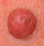
Nevus cell nevus
| Classification according to ICD-10 | |
|---|---|
| D22.9 | Nevus cell nevus |
| ICD-10 online (WHO version 2019) | |
A nevus cell nevus (abbreviation: NZN ; from Latin “naevus” = birthmark) is a brown-colored, sharply delimited patch of skin that is congenital or after birth and consists of nevus cells. It is a particular subtype of pigmented, benign, localized abnormalities of the skin ( pigmented nevi ), commonly known as "moles" or " liver spots ".
histology
Nevus cell nevi consist of nevus cells , a type of cell that is very similar to melanocytes and can also produce brown pigment ( melanin ). However, they differ from melanocytes in their spherical to spindle-shaped shape and arrangement in typical nests. In addition, they can no longer release their pigment to the surrounding skin cells because they lack the dendrites required for this. The deeper they sink into the dermis, the more likely they are to lose their ability to synthesize melanin. Like melanocytes, they come from the neural crest in the skin.
Classification and development
Nevus cell nevi are acquired at a young age (they do not exist since birth, but only appear in the course of life) and undergo a typical development. It is not absolutely necessary to go through all stages; at the end of its development, the nevus cell nevus usually regresses completely (with the exception of dermal nevi in late adulthood).
According to their location in the layers of the skin, the nevus cell nevi are divided into the following three subspecies (according to the stages of their development):
Junction nevus
In the first stage, a NZN grows exactly at the border between epidermis and dermis (the so-called junction zone ) to a sharply delimited, punctiform, brown or black skin change. The macroscopic appearance hardly differs from a lentigo simplex , which, however, consists only of melanocytes and not of nevus cells . Lentigo simplex and junction nevus are the classic " liver spots ". The first junction nevi arise in childhood .
Compound nevus
In the next stage, the NZN migrates a little deeper into the dermis and thus expands into both skin layers ( epidermis and dermis ), whereby the entire change becomes thicker and has more raised and nodular parts. The surface of the lesion may appear fissured. The pigmentation is usually a little more irregular and lighter. The transition to compound nevi usually takes place during puberty . Since the compound nevus is an intermediate phase, it is also called an intermediate nevus .
Dermal nevus
In the final stage of development, the NZN takes on a large, round, hemispherical shape. Its nevus cells have completely sunk into the dermis . Most of the time it has lost its brown pigment and has some hair ( hypertrichosis ). Telangiectasia can develop at the edge of the lesion . Towards the end of the development there is an increasing connective tissue transformation. The large, cosmetically disturbing dermal nevi typically appear in early adulthood .
Other kinds
There are various special forms of nevus cell nevi:
- Congenital nevus cell nevus (large NZN that are already present at birth)
- Halonevus (NZN with depigmented halo)
- Spitz nevus (nodular NZN in children and adolescents)
In addition, under certain circumstances, nevus cell nevi can develop cell atypia and undergo dysplastic changes and thus become dysplastic nevi (presumably precursors of malignant melanoma ).
Clinical significance
etiology
NZN are based on developmental disorders in the embryonic stage and arise from postzygotic mutations in the genetic make-up; however, skin changes only develop in the course of life. The importance of sun exposure is unclear.
Epidemiology
Due to the stages of development, as well as the emergence and disappearance of NZN, a white-skinned adult has an average of about 20 acquired nevus cell nevi. Very light skin types tend to have an increased incidence of NZN. They are less common in dark-skinned people. Ordinary NZN only form in the first half of life and usually disappear completely in the elderly.
clinic
NZN are usually harmless and asymptomatic , but there is a risk of dysplasia and malignant degeneration , which can be noticeable as an increase in size, change in color, pain or itching.
therapy
NZN, which are cosmetically disruptive, can be superficially removed by means of electrocautery , provided that it is guaranteed benign .
For an unequivocal decision about the dignity of an NZN (in the case of unusual localization or abnormalities according to the ABCDE rule ), the lesion and deeper tissue layers must be excised .
forecast
In general, normal nevus cell nevi have a good prognosis. It is important to recognize the potential progression to dysplastic nevi , as malignant melanomas can develop from them . The melanoma risk is directly related to the total number of NZN.
In the event of bleeding, itching, increased size, color changes or other abnormalities (see ABCDE rule ), it is recommended to consult a dermatologist.
literature
- Thomas B. Fitzpatrick, Klaus Wolff (ed.): Atlas and synopsis of clinical dermatology: common and threatening diseases . 3. Edition. McGraw-Hill, New York; Frankfurt a. M. 1998, ISBN 0-07-709988-5 .
- Ernst G. Jung, Ingrid Moll (Ed.): Dermatology . 5th edition. Thieme, Stuttgart 2003, ISBN 3-13-126685-6 .


