Cryoelectron microscopy
The cryo-electron microscopy (cryo-EM) is a form of transmission electron microscopy (TEM), in which biological samples at cryogenic be examined temperatures (≲ -150 ° C).
principle
With conventional electron microscopy of a biological sample at room temperature, the water must be removed from the sample before the examination. The sample has to go through a lengthy and error-prone process in which it is treated with various sample preparations and the water it contains is successively replaced by a plastic embedding medium . In the course of this treatment, artifacts (structural changes) can form in the sample , which can ultimately falsify the result or lead to misinterpretations.
The cryoelectron microscopy enables an image of the examined object close to the native state . Therefore, in comparison to classic TEM, the use of fixation and contrast media can be dispensed with. Without the contrast agent, biological samples are almost transparent to the radiation used due to the uniform, low electron density . Due to the high proportion of ice, only a short dose of radiation intensity (below 1 to 10 electrons per Å 2 ) can be used. As a result, the signal-to-noise ratio is comparatively low. By calculating the structures from thousands of recordings of the same object from different perspectives, a 3D model of the object can be created ( single particle method ). Further improvements can be achieved by averaging several 3D models of the same object ( subtomogram averaging "sub- tomogram averaging "). The resolution of cryoelectron microscopy is usually somewhere between transmission electron microscopy and X-ray structural analysis . In some cases, resolutions around 0.2 nm (2 Å) can be achieved. It is also possible to make three-dimensional recordings of larger structures using cryoelectron tomography .
The cryoelectron microscope
Cryo electron microscopes are specially adapted transmission electron microscopes. Most transmission electron microscopes are equipped with a cold trap . This protects the sample from contamination during exposure to the electron beam and improves the fine vacuum in the area of the sample. The combination of an extremely powerful cold trap (in particular so-called cryoboxes) with a cooled sample holder, which has a thermally connected tank for liquid nitrogen, results in a cryoelectron microscope. Due to the increased use of the research method since 2005, the leading manufacturers of transmission electron microscopes are also building dedicated cryo electron microscopes. By using a direct CMOS recording, images can still be recorded despite the low radiation intensity. The contrast of the image can be enhanced by using phase plates and energy filters .
method
Sample preparation
In cryoelectron microscopy, the sample is shock-frozen ultra- quickly or, more precisely, formulated: vitrified , that is, it is converted into an amorphous, glass-like state. Typically cooling rates of> 10,000 K / s can be achieved. Existing water solidifies to amorphous ice . In particular, cryoelectron microscopes are used to analyze complex protein structures. The structures are cooled to temperatures below −150 ° C (123 K) within fractions of a second using cryogenic liquids such as liquid nitrogen , liquid helium or, preferably, liquid ethane .
Data analysis
The specimen is analyzed from different angles via an adjustment device built into the device.
Data processing
A three-dimensional electron density can be calculated from the data obtained with the aid of computer programs. The basic programs were largely co-developed by Joachim Frank . In further steps, atomic 3-D models of the corresponding biomolecules can then be incorporated into the electron density, similar to X-ray structure analysis.
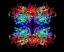
The resolution of 3-D structures determined by cryoelectron microscopy can currently reach 0.2 nanometers (2 Å). The highest resolution so far has been obtained for a glutamate dehydrogenase (GDH) of 1.8 Å. In contrast to X-ray structure analysis, there is a lower theoretical limit for the molecular size of the cryo-EM method, which is around 30 kDa. Another problem with some cryo-EM structures is the different resolution of the electron density within a biomolecule.
variants
Various techniques and variants for cryoelectron microscopy have been developed, e.g. B. electron crystallography , single particle analysis , cryoelectron tomography , MicroED and time-resolved cryo-EM.
history
Cryoelectron microscopy was developed by the Swiss chemist Jacques Dubochet at the European Molecular Biology Laboratory and further developed by Joachim Frank and Richard Henderson . In 2017, all three were awarded the Nobel Prize in Chemistry for their work .
A study on the characterization of Mycobacterium smegmatis shows a comparison of different electron microscopic working techniques with cryolectron microscopy .
literature
- Kira Welter, Cryo-Electron Microscopy: Cool Pictures in 3D Chemistry in Our Time Vol. 6, 2017, pp. 366–368 doi : 10.1002 / ciuz.201770604
Individual evidence
- ↑ a b c d e f g h Maria Mulisch: Romeis - microscopic technology. Springer-Verlag, 2015, ISBN 978-3-642-55190-1 , p. 169.
- ↑ Resolution advances in cryo-EM enable application to drug discovery. In: Curr Opin Struct Biol. Volume 41, Dec 2016, pp. 194-202. doi: 10.1016 / j.sbi.2016.07.009 .
- ↑ J. Vonck, DN Parcej, DJ Mills: Structure of Alcohol Oxidase from Pichia pastoris by Cryo-Electron Microscopy. In: PloS one. Volume 11, number 7, 2016, p. E0159476, doi : 10.1371 / journal.pone.0159476 , PMID 27458710 , PMC 4961394 (free full text).
- ↑ Jacques Dubochet et al. Electron microscopy of frozen-hydrated bacteria Journal of Bacteriology, July 1983, p. 381-390 (page 381). Retrieved January 3, 2018
- ^ J. Vonck & DJ Mills: Advances in high-resolution cryo-EM of oligomeric enzymes. In: Curr Opin Struct Biol. Volume 46, Oct 2017, pp. 48-54. doi: 10.1016 / j.sbi.2017.05.016 .
- ↑ Xiaodong Zou: Electron Crystallography. OUP Oxford, 2011, ISBN 978-0-199-58020-0 . P. 4.
- ^ Grant J. Jensen: Cryo-EM Part B: 3-D Reconstruction. In: Methods in Enzymology. Volume 482, Academic Press, 2010, ISBN 978-0-123-84992-2 . P. 211.
- ^ Joachim Frank: Electron Tomography. Springer Science & Business Media, 2008, ISBN 978-0-387-69008-7 , p. 50
- ^ RA Crowther: The Resolution Revolution: Recent Advances In cryoEM. In: Methods in Enzymology , Volume 579, Academic Press, 2016, ISBN 978-0-128-05435-2 , p. 369.
- ↑ Ziao Fu, Sandip Kaledhonkar, Anneli Borg, Ming Sun, Bo Chen, Robert A. Grassucci, Måns Ehrenberg, Joachim Frank: Key Intermediates in Ribosome Recycling Visualized by Time-Resolved Cryoelectron Microscopy . In: Structure . 24, No. 12, 2016, pp. 2092–2101. doi : 10.1016 / j.str.2016.09.014 . PMID 27818103 . PMC 5143168 (free full text).
- ↑ Xiangsong Feng, Ziao Fu, Sandip Kaledhonkar, Yuan Jia, Binita Shah, Amy Jin, Zheng Liu, Ming Sun, Bo Chen, Robert A. Grassucci, Yukun Ren, Hongyuan Jiang, Joachim Frank, Qiao Lin: A Fast and Effective Microfluidic Spraying-Plunging Method for High-Resolution Single-Particle Cryo-EM . In: Structure . 25, No. 4, 2017, pp. 663–670.e3. doi : 10.1016 / j.str.2017.02.005 . PMID 28286002 . PMC 5382802 (free full text).
- ↑ Bo Chen, Sandip Kaledhonkar, Ming Sun, Bingxin Shen, Zonghuan Lu, David Barnard, Toh-Ming Lu, Ruben L. Gonzalez, Joachim Frank: Structural Dynamics of Ribosome Subunit Association Studied by Mixing-Spraying Time-Resolved Cryogenic Electron Microscopy . In: Structure . 23, No. 6, 2015, pp. 1097-105. doi : 10.1016 / j.str . 2015.04.007 . PMID 26004440 . PMC 4456197 (free full text).
- ↑ Information from the Nobel Foundation on the 2017 award ceremony to Jacques Dubochet, Joachim Frank, Richard Henderson (English)
- ↑ CK Bleck, A. Merz, MG Gutierrez, P. Walther, J. Dubochet, B. Zuber, G. Griffiths: Comparison of different methods for thin section EM analysis of Mycobacterium smegmatis. In: J Microsc. Volume 237, No. 1, January 2010, pp. 23-38, PMID 20055916


