Pityriasis versicolor
| Classification according to ICD-10 | |
|---|---|
| B36.0 | Pityriasis versicolor |
| ICD-10 online (WHO version 2019) | |

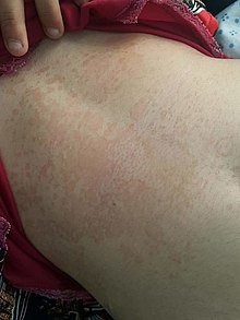
Pityriasis versicolor (PV), also known as bran fungus , is a common, non-contagious, recurrent fungal infection of the upper layer of the skin ( epidermis ), which manifests itself as a map-like dark discoloration. In the temperate latitudes, PV is found in around 1–4% of the population, with an increased incidence during the summer months. In the tropics it can be found in up to 50% of the population.
PV is caused by the yeast Malassezia furfur , which is part of the normal skin flora of almost 100% of the population and mainly feeds on sebum. As a yeast, the fungus remains in the unicellular stage, so it does not develop any fruiting bodies or mycelium . In the case of bran fungus, the affected areas of the skin become overgrowth with the fungus, which produces a brown pigment. The discoloration occurs mainly in the areas rich in sebum glands and thus in the chest, upper back and face. The reasons why the fungi become pathogenic (pathological) in some people are not entirely clear. However, it has been observed that skin mycosis occurs more intensely in the summer months and in people who tend to sweat profusely ( hyperhidrosis ) and increased sebum production ( seborrhea ).
If there is exposure to sunlight, healthy skin tans. Those areas that are affected by the bran fungus, on the other hand, are shielded from UV radiation by the pigments of the fungus. In addition, the Malassezia yeasts that cause it produce a substance that inhibits melanin production by melanocytes . Fine, scaly, red-brownish macules form, preferably on the trunk. Light, whitish macules usually appear on tanned skin. This is where the addition “versicolor” comes from, which in Latin means “color rotation”.
The diagnosis is usually made by the dermatologist as a visual diagnosis and can be confirmed by scraping off flakes of skin (or removing it with an adhesive strip) and examining it under the microscope. The fungal cells can be seen as grape-shaped globules. Important to anamnestic rule out other Dermatomycoses is the only minor itching, which in some heat can be stronger.
Bran fungus does not cause any health restrictions and is therefore viewed more as a cosmetic problem. A high level of relapse is observed in people with a strong propensity for P. versicolor .
Meaning of the Malassezia yeast
Malassezia yeasts belong to the resident skin flora of all people . They are mainly found in areas of the skin that are rich in sebum glands, because their lipophilicity means that they are dependent on the supply of longer-chain fatty acids. A predominance of Malassezia furfur has been demonstrated in lesions of PV . Scientific in vitro studies have also shown that M. furfur is able to synthesize brown pigments when tryptophan is the sole nitrogen source. The compounds that are formed in the process and are hitherto unknown have interesting biological effects which could be related to the pathogenesis of PV; u. a. diagnostically usable fluorescence ( Wood light ), UV protection or induction of apoptosis in human melanocytes. Interestingly, a single enzyme is responsible for the synthesis of the multitude of complex compounds - the so-called tryptophan aminotransferase Tam1.
Symptoms
The skin changes are mostly located in areas with high sebum production, preferably on the chest, back and neck. Sharply demarcated, red-brownish, lenticular to cent-sized macules (Pityriasis versicolor rubra) develop, which can slowly confluence to larger, polycyclic spots. Fine scaling of the herd is typical. If you stroke the lesions with a wooden spatula, the loosened horny layer is lifted out finely (= pityriasiform) ("wood chip phenomenon").
Model for the pathogenesis of Pityriasis versicolor
The research results suggest that tryptophan- dependent compounds synthesized by Malassezia yeasts are important in the pathogenesis of PV , which are induced in the skin environment in the absence of other usable amino nitrogen compounds.
The following model for the pathogenesis of PV results from these findings:
- As a normal component of the resident skin microflora, Malassezia yeasts initially metabolize readily available nitrogen sources such as B. glycine , alanine or serine .
- Under favorable conditions, such as high humidity or increased sebum production, the fungus grows more intensely and thus consumes easily available nitrogen sources.
- After amino acids that are easy to metabolize have been used up, the less preferred amino acid tryptophan is used as a nitrogen source. With the help of the enzyme tryptophan aminotransferase 1 (Tam1), tryptophan is metabolized to a large number of biologically active compounds (indole derivatives).
- The resulting biologically active compounds are responsible for the characteristic picture of pityriasis versicolor, such as the brown discoloration on the skin or the fluorescence used for diagnostics under Wood light .
In view of this model, the hyperpigmentation in the case of PV, in contrast to other forms of hyperpigmentation, appears independent of melanin synthesis. This is supported by the unchanged number of melanosomes in lesions of PV, but also by the occurrence of hyperpigmented areas in areas of vitiligo .

treatment
Conventional treatment is mainly external with prescription antimycotics ( clotrimazole , econazole , bifonazole , sertaconazole , terbinafine or naftifine ), in severe cases also with systemic (internal) therapy with fluconazole , itraconazole or ketoconazole (all subject to prescription). In addition, in some cases, the use of an active ingredient-containing shampoo can be recommended. The irregular and varied pigmentation of the skin can persist for several months after successful treatment. A high relapse rate has been observed in people with a strong propensity for P. versicolor .
Alternatively, an antifungal-free cosmetic application for the dark appearance of pityriasis versicolor is available without a prescription. The "active ingredient" is not specified by the manufacturer, it is supposed to inhibit the mushroom's own enzyme, which is responsible for the production of the brown pigment. There is no evidence of the mechanism of action.
A study from the Jundishapur Journal of Microbiology describes the in vitro use of extracts from the henna (Lawsonia inermis) as a fungicide in Malassezia cultures that exactly Malassezia type is not specified in the study. Overall, however, the effect of the henna extract was inferior to that of miconazole. Shampoos with henna extract are sometimes advertised as an anti-pityriasis agent, but there is no evidence of the in-vivo effect.
Web links
Individual evidence
- ↑ Pityriasis versicolor (overview) B36.0 , from enzyklopaedie-dermatologie.de , accessed on September 17, 2019
- ↑ Mayser PA, Preuss J. Dermatologist 2012: 63: 859-867.
- ↑ Lee WJ, Kim JY, Song CH et al. (2011) Disruption of barrier function in dermatophytosis and pityriasis versicolor . J Dermatol 38 (11): 1049-1053
- ↑ Mostafa WZ, Assaf MI, Ameen IA et al. (2012) Hair loss in pityriasis versicolor lesions: a descriptive clinicopathological study . J Am Acad Dermatol
- ↑ Epicolor Body Fluid , from www.epicolor.de, accessed on September 17, 2019
- ↑ Berenji: In vitro study of the effects of Henna extracts (Lawsonia inermis) on Malassezia species. Retrieved December 14, 2019 .
