Primitive stripes
In developmental biology, primitive streak is the name for a bead of cells that temporarily forms on the germ or germinal disc of reptiles , birds and mammals in early embryogenesis .

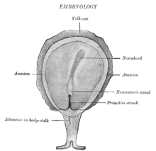
The photo shows a
- above the allantois in the stalk , at the caudal end of the embryos in the direction of neurenteric canal extending -
primitive streak "Primitive streak" with primitive groove and primitive pit (which the primitive node containing).
The two-layer embryonic disc shows on the dorsal germinal layer ( epiblast ) bordering the amniotic cavity , initially as a result of proliferation, a displacement of cells that accumulate from two sides in the midline of the caudal area and thus represent the primitive streak , in humans around the 17th day of its development. At this stage, the embryo visibly gains an orientation on a rostro-caudal longitudinal axis, which allows it to be distinguished from caudal (posterior, back) to rostral (anterior, front) on two sides in halves. The primitive streak then becomes stronger due to immigrating cells (migration) and longer in the caudal direction. It sinks into a primitive channel, the rostral region of which is reinforced by cell immigration and becomes a primitive pit .
The primitive stripe and the primitive knot located in the marginal wall of the primitive pit are of decisive importance in the organization of spatial patterns in the development control of an embryo ( morphogenetic organization). In amniotic vertebrates, the depression of the primitive stripe to the primitive trough represents the beginning gastrulation . The primitive knot , also called Hensian knot in birds (after Victor Hensen ), is a knot-like swelling in the wall of the edge area of the primitive pit in the sauropsid and mammalian germ . Functionally, the knot corresponds to the Spemann organizer on the dorsal lip of the ancient mouth of amphibians .
(Nodal) cells located in the area of the primitive node generate a flow (clockwise from the ventral view) with the primary monocilia , which is inclined posteriorly and rotates - chirally composed of microtubules and specific dynein - in the overlying extraembryonic fluid, via which signal substances are distributed. These morphogens are distributed by the left-hand flow, and the cells sensitive to these signals are addressed to different degrees as a result of the gradient. This leads to an asymmetrical cell pattern, for example in the expression of Nodal . This becomes the reason for the lateralization of prospective internal organs that later emerge from this cell layer, such as the heart that is on the left . If the direction of flow is reversed, the situation is reversed; a situs inversus can develop in humans , e.g. B. in the context of a Kartagener syndrome , caused by a disturbed motility of (nodal) cilia of the primitive node.
Individual evidence
- ↑ W. Janning, E. Knust: Genetics . Thieme, Stuttgart 2008, ISBN 978-3-13-128772-4 , Chapter 30.2 (Formation of the left-right asymmetry), p. 463.