
Tympanometry
In the Tympanometry is an objective measurement method of Audiology . It belongs to the impedance audiometry. In this case, the impedance is understood to mean the extent of resistance that the acoustic system as a whole, e.g. B. the middle ear, opposed to the reception of sound waves . An acoustic system with high impedance absorbs little sound energy and reflects a large part. In contrast, a system with low impedance absorbs a lot of sound energy in the form of vibrations . The extent of sound absorption is also described here by the term “compliance” and means the flexibility or rigidity of the eardrum. The aim is to indirectly measure the pressure in the middle ear by measuring the acoustic resistance of the eardrum, to detect pathological middle ear contents and to assess the condition of the ossicular chain . The measurement method is based on the work of Schuster (1934), Metz (1946), Zwislocki (1957) and Terkildsen and Nielsen (1960). The stapedius reflex is often co-determined with the measurement system.
Measuring principle
With tympanometry, a pressure fluctuation is created in the ear canal. A negative pressure is followed by a slight overpressure. These changes in pressure are reflected by the eardrum and then measured using a sealed probe. Tympanometry is a practical, standardized diagnostic procedure for examining the eardrum and middle ear. The measurement parameter in tympanometry is the flexibility or stiffness of the eardrum (so-called eardrum compliance), the reciprocal of which is the impedance.

functionality
When the eardrum is exposed to sound with a defined amount of sound energy , part of this energy is reflected and the remaining part is passed on to the middle ear. The amount of reflected energy depends on the acoustic resistance, the acoustic impedance of the eardrum. Under natural pressure conditions, when there is the same atmospheric pressure in front of and behind the eardrum, the transmission of the sound is best due to the physical properties of the eardrum.
The prerequisite for performing this test is an intact eardrum and an airtight closure of the ear canal by the measuring probe.

A separate device, a tympanometer, is required to perform tympanometry. The measurement is carried out using a measuring probe that has three bores with small tubes. A continuous tone (“probe tone”) with a frequency of mostly 226 Hz (95 dB SPL ) is given through the first hole by a tone generator . The second hole contains a microphone that is connected to a measuring instrument. A pressure pump is used to build up defined pressures in the ear canal via the third hole. Under normal conditions, i.e. when the pressure conditions in the auditory canal and middle ear are the same and the eardrum is in the "normal position", there is a certain acoustic resistance which is used by the tympanometer as a reference point (zero point). If an overpressure or underpressure is now generated by the pressure pump, the eardrum is tensioned and the acoustic resistance of the eardrum changes. This also increases the amount of reflected sound energy of the probe tone, the sound pressure level in the ear canal increases, which can be determined by the connected measurement microphone. The changed reflections are shown graphically in the tympanogram as compliance of the eardrum (elasticity, reciprocal value of the acoustic resistance).
Due to the much smaller auditory canal volumes in infants, the use of a probe tone of 226 Hz leads to falsified measurement results, so a 1000 Hz probe tone should be used instead.
Clinical application
Clinical applications are e.g. B. the following diseases or issues:
- Otitis media
- Perforation of the eardrum
- Dysfunction of the auditory ossicles
- Eustachian tube malfunction
- Otosclerosis
- Tympanosclerosis
- Cholesteatoma
Clinical evaluation
Depending on the clinical picture, the normal curve changes in the tympanogram, which can be used to identify the disease. With normal middle ear function, the maximum mobility of the eardrum is around 0 Pa , in other words the eardrum can swing out to the maximum, since the same pressure prevails in the external auditory canal as in the tympanic cavity . The measured curve is referred to as the A curve, see Figure I. If there is a negative pressure in the tympanic cavity, which may be the result of a tube ventilation disorder, the measurement of such patients results in a shift of the maximum into the negative pressure range, see Figure II. The curve measured is called the C curve.
A middle ear, caused for example by an acute otitis media , acute otitis media typically results in the measurement to a flat tympanogram, see Figure III. This is due to a build-up of fluid in the middle ear. The measured curve is called the B curve. An interruption in the sound conduction chain, for example due to an ossicular dislocation but also due to atrophic eardrum scars , leads to an upwardly open curve , not shown.
With a tube catarrh z. B. the eardrum is pulled inward due to the disease, retracted. If a negative pressure is now generated in the auditory canal via the pressure pump, the eardrum returns to its normal position and normal compliance is achieved. The measured values are entered in the tympanogram form and show a shift in the negative pressure range.
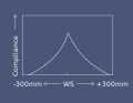
I. Normal tympanogram, maximum at 0 Pa.
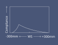
II. Negative pressure in the middle ear (e.g. tubular catarrh ), maximum in the negative pressure range

III. Fluid in the middle ear (e.g., tympanic effusion )


The vertical or ordinate axis of the tympanogram provides information about the compliance of the middle ear space , more precisely the mobility of the eardrum in ml or in relative volume units . The horizontal or x-axis shows the corresponding change in pressure in Pa or decaPascals (1 daPa = 10 Pascal = 0.1 m bar ) to, by some equipment manufacturers, the pressure data are also in mm H 2 O is applied.
Individual evidence
- ^ Rudolf Probst; Gerhard Grevers; Heinrich Iro: ear, nose and throat medicine. Georg Thieme, Stuttgart 2000, ISBN 3-13-119031-0 , p. 184
- ↑ Schuster, K .: A method for comparing acoustic impedances . In: Physikalische Zeitschrift . tape 35 , 1934, pp. 408-409 .
- ^ Metz, O .: The acoustic impedance measured on normal and pathological ears . In: Acta Oto-Laryngologica Supplement . tape 63 , 1946, ISSN 0365-5237 , p. 1-254 .
- ↑ Zwislocki, J .: Some measurements of the impedance at the eardrum . In: Journal of the Acoustical Society of America . tape 29 , 1957, pp. 349-356 , doi : 10.1121 / 1.1908887 .
- ↑ Terkildsen, K. and Nielsen, S .: An electroacoustic impedance measuring bridge for clinical use . In: Archives of Otolaryngology . tape 72 , 1960, pp. 339–346 , doi : 10.1001 / archotol.1960.00740010347009 .
- ↑ Kun Yang, Zhiqi Liu: Comparison of 1000 Hz-, 226 Hz-probe tone tympanometry and magnetic resonance imaging in evaluating the function of middle ear in infants . In: International Journal of Pediatric Otorhinolaryngology . tape 136 , no. 9 , 2020 ( [1] [accessed August 12, 2020]).