Balance organ

In the organ of equilibrium of living beings, various sensors are used to perceive linear accelerations (including gravitational acceleration ) and angular accelerations . The stimulus is usually absorbed by sensory cells that are coupled to one or - as in humans - several specially suspended or resting solid bodies, so-called statoliths . A liquid in a pipe system is often used as an inertial mass for the rotary movements. In all vertebrates, including humans , the vestibular apparatus is the most important organ of equilibrium.
The vestibular apparatus of the vertebrates

1 vestibular nerve
2 cochlear nerve
3 facial nerve
4 external facial knee with Ggl. geniculi
5 chorda tympani
6 cochlea
7 semicircular canals
8 hammer handle
9 eardrum
10 eustachian tube
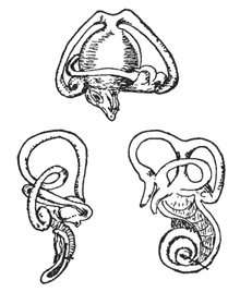
The paired vestibular organ ( organon vestibular , vestibular apparatus) of vertebrates and humans is located in the inner ear . It is usually divided into five components each: three semicircular canals and the two structures called macular organs sacculus ( Latin : 'sac') and utriculus (Latin: 'small tube'). Fish and amphibians (see below ) have an additional sixth component, a lagena (Latin: 'bottle'), which is also a macular organ. There are also exceptions to the number of semicircular canals, but only in very primitive vertebrates. Lampreys have only two pairs of semicircular canals, and hagfish only have one pair.
The human vestibular apparatus
Semicircular canal organs
The semicircular canals, which are filled with endolymph (not with air, as was generally assumed before the description by the anatomist Cotugno) form the sense of rotation and are almost perpendicular to each other and thus record the vector components of the rotational accelerations of the head in space. They each consist of the actual arch and an extension, the ampoule . It contains the hair cells of the semicircular canals, the sensory cells of the organ of equilibrium. Their hair protrudes into a gel cone, the cupula , which interrupts the liquid ring. When the head accelerates, the semicircular canals rotate with it. The endolymph, as a liquid, can more or less evade this rotary movement due to its inertia, if the semicircular canal is aligned accordingly. Through this relative movement of the endolymph in relation to the semicircular canal, the endolymph pushes the cupula aside. This causes the "hair" of the sensory hair cells to be bent. Depending on the direction of the turn, there is an acceleration or deceleration of the resting frequency of the sensory hair cells. The electrical signals reach the brain via the semicircular canal nerve.
Macular organs
Sacculus and utriculus record the translational acceleration of the body in space. They are also perpendicular to each other so that the saccule responds to vertical and the utriculus to horizontal accelerations. The sensory cells protrude with their appendages (sensory hairs, especially stereocilia ) into a gelatinous membrane that contains otoliths (statoliths). Otoliths are fine calcium carbonate crystals , which increase the density of the membrane and thus in turn enable an inertia effect, so that the detection of linear accelerations is made possible at all.
Processing in the nervous system
The sensory information arrives from the sensory cells via the 8th cranial nerve ( vestibulocochlear nerve ) to corresponding nerve nuclei in the brain stem ( vestibular nuclei ). These receive additional information from the eyes, the cerebellum, and the spinal cord .
The interconnection of the organ of equilibrium with the eye muscles ( vestibular ocular reflex ) enables the visual perception of a stable image while the head is moving at the same time.
In addition to the equilibrium system ( vestibular system ), the visual system and the proprioceptive system ( depth sensitivity ) are also responsible for conscious orientation in space .
If the function of one of these systems is disturbed, this can result in contradicting information from the individual sensory organs. This can make you feel dizzy . Malfunction of the otoliths can cause benign positional vertigo.
Recent studies show that the organ of equilibrium in the inner ear is not only responsible for spatial orientation: it also plays an important role in the precise control of body movements. This function seems to play an important role, especially for movements in the dark or for complex sequences of movements, such as those performed by gymnasts or artists .
Balance check
Coordination tests
- Romberg experiment : The examined person stands with his eyes closed so that the feet touch each other inside. The arms are stretched out horizontally. The examiner assesses the subject's stability or tendency to fall.
- Unterberger-Tretversuch : The examined marches with closed eyes "in one place", if necessary with the arms stretched forward. The examiner assesses the deviation to the right or left.
- Gait deviation : When walking forward with closed eyes, the gait deviation is assessed.
- Berg Balance Scale , a test procedure in which the balance behavior and the "risk of falling" are determined on the basis of 14 tests.
Experimental tests
- Caloric examination of the organ of equilibrium : During the examination, the patient lies on his back with his head slightly raised. Your eyes should be closed so that orientation in the room is not possible. Rinsing the ear canal with cold or warm water (30 ° C, 44 ° C) causes movement of the endolymph in the vestibular organ, which is associated with dizziness. If the vestibular organ is intact, nystagmus , i.e. a typical lateral twitching of the eye, can be observed and evaluated. As a rule, the eye moves in the direction of the irritated ear with a warm rinse, and in the opposite direction with a cold stimulus. If the eardrum is not intact, do not rinse with water. Alternatively, the experiment can be carried out with diethyl ether or with air.
The vestibular apparatus of fish and amphibians
In addition to the semicircular canals, all fish have three macular organs, each of which contains an otolith . The saccule in particular serves the sense of hearing , the differences in density between the sagitta and the surrounding endolymph in the case of sound waves in the near field leading to shear movements in the hair cells. To expand the sense of hearing to greater distances and higher frequencies, some bony fish species have special coupling mechanisms between their swim bladder and the skull bone or their inner ear. In a few cases, the inner ear is surrounded by special air-filled bubbles.
| Macular organ | Name of the otolith | function | variability | relative size |
|---|---|---|---|---|
| utricle | Lapillus | Acquisition of horizontal linear accelerations | low | mostly small |
| Saccule | Sagitta | Acquisition of vertical linear accelerations | large, with not one of the Ostariophysi belonging bony fish | large, extremely large (over 30 mm) in umberfishing |
| Lagena | Asteriscus | Hear and capture vertical linear accelerations | large, especially among the Ostariophysi | medium |
Amphibians also have a Lagena, which, however, only perceives acceleration. As far as is known so far, the sacculus of these animals is only used to perceive substrate vibrations, while the amphibiorum papilla can also pick up sound and the basilar papilla is used exclusively for hearing.
Other organs of balance
The organs of equilibrium in birds
Birds even have several independent organs of balance. They have a second organ of equilibrium in the lateral flaps of the spinal cord . It is solely responsible for controlling walking and standing. The vestibular apparatus in the inner ear, on the other hand, controls the movements of birds in flight .
The organs of equilibrium in insects
A large number of organs of insects have been described that presumably or have been proven to serve as an organ of equilibrium:
- the swinging bulb ,
- Graber's organ in the abdomen of horsefly larvae ,
- the palm organ in the head of mayflies (static sense demonstrated in larvae) and
- the statocysts on the 10th and 11th abdominal segment of the larvae of a wrinkle mosquito .
Other animals
In the animal kingdom, organs of equilibrium with a kinetically freely moving solid, a statolith , which consists of the body's own material and which has arisen through biomineralization within the body or has been taken in from outside, are widespread . Such organs are usually called statocysts and can be found, for example, in:
- Rib jellyfish ,
- Planarians ,
- Molluscs ,
- some annelids and
- some crustaceans .
Since the statoliths of crayfish are located in pits at the base of the first pair of antennae, they are lost when they molt and must be replaced by the animals with a pebble from their surroundings. This fact formed the basis for experiments in which only iron granules were made available to the crabs after molting. The static sense could be disrupted and specifically examined with the help of artificial magnetic fields.
See also
- Sense of balance
- Semicircular canals
- Nystagmus
- Vestibular evoked myogenic potentials
- Inertial navigation system
- Vestibular syndrome
Web links
- Movement in balance. On: Wissenschaft.de , August 9, 2005. The organ of equilibrium in the inner ear coordinates complex motor sequences.
Individual evidence
- ↑ Christopher Platt, Arthur N. Popper: Fine structure and function of the ear. In: William N. Tavolga, Arthur N. Popper, Richard R. Fay (Eds.): Hearing and sound communication in fishes. Springer, New York NY a. a. 1981, ISBN 0-387-90590-1 , pp. 3-38, doi : 10.1007 / 978-1-4615-7186-5_1 .
- ^ Domenico Cotugno: De aquaeductibus auris humanae internae. Anatomica Dissertatio. Simoniana, Naples 1761.
- ^ Brian L. Day, Raymond F. Reynolds: Vestibular response shapes voluntary movement. In: Current Biology. Volume 15, No. 15, 2005, pp. 1390-1394, PMID 16085491 , doi: 10.1016 / j.cub.2005.06.036 .
- ↑ Arthur N. Popper: Organization of the inner ear and auditory processing. In: R. Glenn Northcutt, Roger E. Davis (Eds.): Fish Neurobiology. Volume 1: Brain stem and sense organs. The University of Michigan Press, Ann Arbor MI 1983, ISBN 0-472-10005-X , pp. 126-178.
- ↑ Stefan Holler: Convergence of afferent and commissural signals from the semicircular canals and the otolith organs in the common frog (Rana temporaria). Munich 2001, university, dissertation; Digitized version (PDF; 4.93 MB).
- ^ Necker, R. (2005). The structure and development of avian lumbosacral specializations of the vertebral canal and the spinal cord with special reference to a possible function as a sense organ of equilibrium. Anat. Embryol., 210 (1): 59-74. doi: 10.1007 / s00429-005-0016-6
- ^ Necker, R. (2006). Specializations in the lumbosacral vertebral canal and spinal cord of birds: evidence of a function as a sense organ which is involved in the control of walking. J. Comp. Physiol .: A-Sens. Neur. Behav. Phys., 192 (5): 439-448. doi: 10.1007 / s00359-006-0105-x
- ↑ Necker, R., Janßen, A., and Beissenhirtz, T. (2000). Behavioral evidence of the role of lumbosacral anatomical specializations in pigeons in maintaining balance during terrestrial locomotion. J. Comp. Physiol .: A-Sens. Neur. Behav. Phys., 186 (4): 409-412. doi: 10.1007 / s003590050440
- ↑ Rolf Gattermann (Ed.): Dictionary of the behavioral biology of animals and humans. 2nd, completely revised edition. Elsevier - Spectrum, Akademischer Verlag, Munich 2006, ISBN 3-8274-1703-1 .


