Vestibular evoked myogenic potentials
Vestibular evoked potentials Myogenic ( VEMP ; engl. : Vestibular Evoked Myogenic potential ) are formed by a reflection of the vestibular system to vibration or acoustic stimuli of the equilibrium organs induced potential differences applied to muscles can be derived. Above all, they serve the selective and side-specific function determination of the saccule ("sac"), one of the organs of equilibrium.

history
The first description of the VEMP from 1992 comes from the Australian researchers Colebatch and Halmagyi. In humans, they were able to display and assign evoked potentials in the electromyogram (EMG) of neck muscles caused by stimulation of the sensory organs of the vestibular system .
Over a decade later, in 2004, another research team also presented myogenic recordings from external eye muscles with supra-threshold acoustic stimulation for the first time . This ocular VEMP (oVEMP) are considered another reflex induced method of examination of the otolith organs .
Basics
To measure VEMP, different types of stimuli are set which , when conducted via air or bones, stimulate the inner ear by causing its membranous labyrinth to vibrate. As a result, sensory cells located at different points of the tube-like structure are addressed and the nerve cells assigned to them are excited. These are, in certain stimuli in addition to those of the hearing sense - in the organ of Corti of the cochlea ( cochlea ) - which also the sense of equilibrium - less in the archway organs than in the macula organs of the vestibular portion.

The signals from the saccule and utricle one side are the lower Vestibularnerventeil to the equilateral vestibular nuclei in the brainstem passed and here switched to other neurons of the vestibular system . These connect via various pathways with other regions in the brain and spinal cord.
About descending pathways including in are cervical spinal cord motor neurons of the throat and neck muscles reached and involved for holding and positioning reflexes, the position changes of the head with tonic answer or phasic muscle activity. A reflex response of the neck muscles can be triggered by stimuli in the labyrinth, which can be measured electromyographically on the muscles close to the surface . The typical stimulus response shows two potential complexes, a “vestibular complex” at 13 and 23 ms, which originates from the saccule, and a “cochlear complex” at 34 and 44 ms.
Motor neurons of the outer eye muscles in the pons and midbrain are reached via ascending pathways and integrated for vestibulo- ocular reflexes that respond to changes in head posture and rapid head movements with compensating eye movements. Therefore, stimuli in the labyrinth, especially the semicircular canal organs, when the head rotates, trigger a reflex response from the eye muscles that interact in the gaze game. However, since the macular organs hardly respond to angular accelerations, but rather to linear accelerations (such as gravitational acceleration ), stimuli that correspond to those of translational movements (such as upwards and downwards) of the head must be set to test them. Under these circumstances, courses can be derived from the corresponding eye muscles in the EMG, which also represent vestibular evoked potentials.
methodology
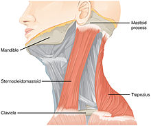
The basis of the VEMP methodology is the sensitivity of certain sensory cells (parastriolar type 1 cells) of the sacculus and utriculus to intense, e.g. Sometimes supra-threshold acoustic stimuli. In clinical practice, derivatives of cervical muscles (cVEMP) such as the sternocleidomastoid muscle for the so-called sacculo-collic reflex and of external eye muscles (oVEMP) such as the rectus superior muscle and the superior obliquus muscle for the so-called utriculo-ocular reflex by means of surfaces -Electromyography (EMG) enforced. The measurements apply to a biphasic muscle potential fluctuation - inhibitory - initially positive potential after 13 ms (p13), then secondary negative potential after 23 ms (n23) for cVEMP - and excitatory stimuli - initially negative potential after 10 ms (n10), then positive potential after 15 ms (p15) for oVEMP - with tonic muscle tension.
stimulation
In daily practice that is currently air line stimulation ( air Conducted sound stimulation , AC or ACS) realized in cervical and ocular discharges. They are carried out using supra-threshold stimulus levels (100 dB nHL) at a stimulus frequency of 500 and 1000 Hz with a sinusoidal burst signal ( tone burst lasting approx. 4–7 ms ). For bone conduction stimuli ( bone conducted vibration , BC or BCV) with bone conduction headphones and transmastoidal acceleration stimuli with a mini-shaker, stimulus frequencies of approx. 100–4000 Hz are effective. There is agreement in international literature that AC-stimulated cVEMPs reflect saccular function in receptor dysfunction. AC- and BC-stimulated oVEMP are considered to be an indicator of utriculus function, but are still discussed controversially.
Despite the non-physiological stimulus of the VEMP, different stimuli (ACS, BCV), different stimulation locations (Fz, Cz, Mastoid), as well as varying stimulus intensity (volume approx. 60–130 dB nHL) at stimulus frequencies of approx. 100–4000 Hz can be achieved carry out an analysis of the otolith function under dynamic aspects.
Diagnostic value
Ocular and cervical VEMP are currently used to diagnose various diseases of the organ of equilibrium. Among other things, this can be used to demonstrate involvement of the sacculus in Menière's disease . VEMPs are also used to precisely determine the extent of nerve inflammation ( neuritis ) of the equilibrium nerve ( vestibular nerve ). The cVEMP and oVEMP findings can be used to determine whether the upper and / or lower part of the equilibrium nerve is involved in the damage in peripheral vestibulopathy.
In neurootology , this examination method represents a supplementary procedure in the context of the equilibrium function test ( equilibriometry ). Clinically, it is used to clarify various issues - in the fields of neurology , otology and ophthalmology - in particular for the objectifiable, laterally separate function test of the two maculae sacculi or the an structures of the brainstem involved in the reflex arcs .
Individual evidence
- ↑ J. Colebatch, G. Halmagyi: Vestibular evoked potentials in human neck muscles before and after unilateral vestibular deafferentation. In: Neurology. Volume 42, No. 8, August 1992, pp. 1635-1636, PMID 1641165 .
- ↑ J. Colebatch, G. Halmagyi: Vestibular evoked myogenic potentials in the sternomastoid muscle are not of lateral canal origin. In: Acta Oto-Laryngologica Supplementum 520. Punkt 1, 1995, pp. 1-3, PMID 8749065 .
- ↑ N. Todd, I. Curthoys, S. Aw, M. Todd, L. McGarvie, S. Rosengren, J. Colebatch, G. Halmagyi: Vestibular evoked ocular responses to air- (AC) and bone-conducted (BC) sound. In: Journal of Vestibular Research. Volume 14, 2004, pp. 123-124 and pp. 215-217, respectively.
- ↑ see video Derivation of the air conduction-induced VEMP at 500 and 1000 Hz ( Memento from May 12, 2014 in the Internet Archive )
- ↑ L. Walther, K. Hörmann, O. Pfaar: Recording cervical and ocular vestibular evoked myogenic potentials. In: ENT. Volume 58, 2010, No. 10, pp. 1031-1045 (Part 1), PMID 20927621 , and No. 11, pp. 1129-1142 (Part 2), PMID 20963394 .
- ↑ L. Walther, I. Repik: neuritis of the vestibular nerve inferior. or Inferior vestibular neuritis: diagnosis using VEMP. In: ENT. Volume 60, 2012, No. 2, pp. 126-131, doi: 10.1007 / s00106-011-2373-1 or PMID 22037927 .
- ↑ G. Zhou, L. Cox: Vestibular evoked myogenic potentials: history and overview. In: American Journal of Audiology. Volume 13, No. 2, 2004, pp. 135-43, PMID 15903139 .
- ^ S. Rauch: Vestibular evoked myogenic potentials. Recording cervical and ocular vestibular evoked myogenic potentials. In: Current Opinion in Otolaryngological & Head and. Neck Surgery. Volume 14, No. 14, 2006, pp. 299-304 PMID 16974141 .
- ↑ Krister Brantberg: Vestibular evoked myogenic potentials (VEMPs): usefulness in clinical neurotology. In: Seminars in Neurology. Volume 29, No. 5, 2009, pp. 541-547, PMID 19834866 .

