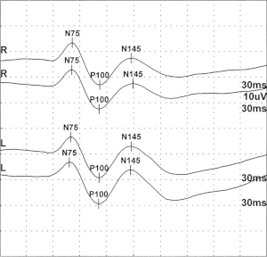
Visually evoked potential
Visual evoked potentials (VEP, also: VECP = v isually e voked c ortical p otentials) are by visual stimulation of the retina induced potential differences less electric charges over the area of the visual cortex may be derived at the back of the skin. They are used in neurology and ophthalmology as a diagnostic procedure and allow an objective detection of the sensory excitation lines up to the cerebral cortex. The measurement of the latency period (transit time) and amplitude (extent of excitability) of the potentials provide information on the function of the visual pathway and its components ( optic nerve , visual cortex). The visual stimulation occurs either with light pulses or with a checkerboard pattern in which the contrasts are reversed at short intervals. In healthy people, the latency period for the primary cortical potential is 100 milliseconds . A negative rash can be seen on the monitor.
In the case of circulatory disorders , degenerative processes and inflammation in the area of the visual pathway, the latency period may be delayed and / or the amplitude may be reduced. VEPs also play an important role in the diagnosis of multiple sclerosis .
Interpretation of the values
A typical VEP has a valley of its potentials at around 80 ms (N80), a peak at 100 ms (so-called P100) and a further minimum at around 135 ms (N135). In the graphic representation, the positive peak P100 is shown pointing downwards (mostly in neurology, see figures) or alternatively pointing upwards (often in ophthalmology and research). The latency of the P100 (recently also called "peak time") and the amplitude, which is measured either as the difference P100-N80 , or as the mean difference ((P100-N80) + (P100-N135)) / 2 are measured . The latency (peak time) is in the broadest sense a value for the time that a light stimulus needs from the retina of the eye as a voltage impulse to the visual cortex in the back of the head. In the normal population it is in the range of 95–115 ms. The amplitude ranges from 5 µV to 30 µV.
The classic application of the VEP is the diagnosis of the visual pathway and its components ( optic nerve , visual cortex ), e.g. in the context of multiple sclerosis. In the case of inflammation in the area of the visual pathway, the latency is significantly increased (by 20 ms and more), the amplitude is slightly reduced. VEPs also allow the severity of amblyopia to be assessed and are therefore used in strabological diagnostics.
- A slight latency (delay) at normal amplitude indicates demyelination. The amplitude can also be reduced here.
- A large delay in the signal at normal amplitude indicates demyelination and remyelination.
- A split and delayed stimulus response indicates chronic de- and remyelination.
- No response indicates complete damage to the axons or there is a conduction block, which may be caused by a tumor.
- A low stimulus response (voltage potential) and a normal latency (delay) indicate partial damage to the axons or a partial conduction block.
The VEP are highly dependent on the technical parameters of the stimulation (monitor, mirror, flash goggles, distance to the eye). It is therefore mandatory that special standard values are determined for the respective constellation. There is also a dependency on age with a slow increase in latency from the age of 55.
literature
- Klaus Poeck , Werner Hacke : Neurology. 12th, updated and expanded edition. Springer, Heidelberg 2006, ISBN 3-540-29997-1 .
- Karl F. Masuhr , Marianne Neumann: Neurology (= dual series ). 4th edition. Hippokrates-Verlag, Stuttgart 1998, ISBN 3-7773-1334-3 .
- Franz Grehn: Ophthalmology. Berlin: Springer Verlag, 30th edition, 2008, ISBN 978-3-540-75264-6
Individual evidence
- ↑ Rudolf Sachsenweger (Ed.): Neuroophthalmology. 3rd, revised edition. Thieme, Stuttgart et al. 1982, ISBN 3-13-531003-5 , p. 51.
- ↑ a b c Michael Bach, University Eye Clinic Freiburg: Ophthalmological electrodiagnostics.
- ↑ Herbert Kaufmann (Ed.): Strabismus. 3rd, fundamentally revised and expanded edition. Georg Thieme, Stuttgart et al. 2004, ISBN 3-13-129723-9 , p. 280.
- ↑ Peter Berlit (Ed.): Clinical Neurology. Springer, Berlin et al. 1999, ISBN 3-540-65281-7 .
- ↑ Klaus Lowitzsch include: the EP-book. Thieme, Stuttgart et al. 2000, ISBN 3-13-116731-9 .


