Tooth root
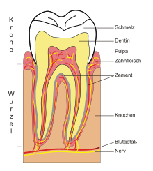
A tooth root ( Latin : Radix dentis ) is the part of a tooth that lies below the tooth crown and secures the tooth in the tooth socket of the jaw . The transition between the tooth crown and the tooth root is the tooth neck . The tooth root consists mainly of the dentin , which is covered by the root cement, which is formed by cementoblasts . In many teeth, a tooth root tapers towards the tip of the root and therefore has a conical shape.
Structure of a tooth root
The tooth pulp , which contains blood vessels and nerve fibers , runs from the pulp cavity in one or more root canals in the direction of the apex dentis and exits through an opening, the foramen apicale dentis , in the alveolar bone.
Permanent teeth
The human teeth have different numbers of roots. The first upper premolar has two roots, the second upper premolar usually only one root. Tooth roots are often partially fused together, or single-rooted teeth separate into two root tips, as can occasionally be observed in upper canines . There is at least one root canal per root. Root anomalies occur. Usually have incisors ( incisors ) and canines ( Canini ), a root with a root canal, the small molars ( bicuspids ) one or two roots with as many root canals. The roots of the wisdom teeth can be very variable, both in number and shape. Some root forms harbor the risk of a root fracture if a tooth extraction is necessary . The complete preparation of the root canals of these teeth is often not possible during root canal treatments.
The point of separation of two roots is called a bifurcation , that of three roots is called a trifurcation .
Number of roots and root canals
The number of tooth roots and root canals of the permanent teeth are shown in the following table, although deviations are possible. The mesial root of the lower molars usually has two root canals. The table shows the percentage frequency of the number of roots (according to Ingle) and the percentage frequency of the number of root canals (according to Schuhmacher).
| teeth | Number of tooth roots | Number of root canals |
|---|---|---|
| upper jaw | ||
| 11, 21 | 1 | 1 |
| 12, 22 | 1 | 1 |
| 13, 23 | 1 | 1 |
| 14, 24 | 1 2 (> 60%) 3 |
1 (9.0%) 2 (85.0%) 3 (6.0%) |
| 15, 25 | 1 (> 85%) 2 |
1 (75.0%) 2 (24.0%) 3 (1.0%) |
| 16, 26 | 3 | 3 (41.1%) 4 (56.5%) 5 (2.4%) |
| 17, 27 | 3 | 3 4 |
| 18, 28 | variable | variable |
| Lower jaw | ||
| 31, 41 | 1 | 1 (70.1%) 2 (29.9%) 3 (0.5%) |
| 32, 42 | 1 | 1 (56.9%) 2 (43.1%) |
| 33, 43 | 1 | 1 (94.0%) 2 (6.0%) |
| 34, 44 | 1 (74.0%) 2 (26.0%) |
1 (73.5%) 2 (26.0%) 3 (0.5%) |
| 35, 45 | 1 (85.0%) 2 (15.0%) |
1 (85.5%) 2 (13.0%) 3 (0.5%) |
| 36, 46 | 2 | 2 (6.7%) 3 (64.4%) 4 (28.9%) |
| 37, 47 | 2 | 2 3 4 |
| 38, 48 | variable | variable |
Milk teeth
Baby teeth, when fully grown, also have tooth roots. The deciduous incisors and canines have one root, the deciduous molars have two roots in the lower jaw and three roots in the upper jaw. Anomalies are very rare in primary dentition. When changing teeth (6 to 12 years of age) with the resulting normal loss of milk teeth, the roots of the milk teeth are resorbed (absorbed) by the permanent teeth that push in and do not seem to have (had) any roots.
Number of milk tooth roots and root canals
The number of tooth roots and root canals of the milk teeth are shown in the following table, although deviations are possible.
| teeth | Number of tooth roots | Number of root canals |
|---|---|---|
| upper jaw | ||
| 51, 61 | 1 | 1 |
| 52, 62 | 1 | 1 |
| 53, 63 | 1 | 1 |
| 54, 64 | 3 | 3 |
| 55, 65 | 3 | 3 |
| Lower jaw | ||
| 71, 81 | 1 | 1 |
| 72, 82 | 1 | 1 |
| 73, 83 | 1 | 1 |
| 74, 84 | 2 | 2 |
| 75, 85 | 2 | 2 |
Artificial tooth roots
Artificial tooth roots are dental implants, after extraction of permanent teeth or aplasia of teeth in the jawbone are planted. A tooth crown is placed on this artificial tooth root or dentures (bridge, prosthesis) are attached to it.
See also
Web links
Individual evidence
- ^ John Ide Ingle, Leif K. Bakland: Endodontics. 4th edition. Williams & Wilkins, Baltimore MD et al. 1994, ISBN 0-683-04310-2 .
- ^ Gert-Horst Schumacher : Anatomy. Textbook and atlas. Volume 1: head, orofacial system, eye, ear, ducts. 2nd, completely revised and enlarged edition. Barth, Leipzig et al. 1991, ISBN 3-335-00264-4 .


