
Extraction (dentistry)

As extraction (from latin extrahere "pull"), exodontia or tooth extraction is used in dentistry of a removal tooth designated without surgery. If an incision and, if necessary, the formation of a periosteal flap of the mucous membrane is required to remove the tooth , this is an osteotomy (surgical tooth removal).
The extraction is usually carried out under local anesthesia in the form of infiltration or conduction anesthesia.
indication


A distinction is made between absolute and relative indications.
Absolute indications
- Severely loosened tooth (degree of loosening III), regeneration of the tooth holding apparatus ( reattachment ) is not to be expected
- Longitudinal fracture of the tooth crown or the tooth root
- Transverse fracture of the tooth root in the middle third of the tooth root
- Massive apical periodontitis when surgical revision ( apicectomy or hemisection ) is not possible
- Lack of space if this cannot be remedied by orthodontic measures alone .
- Removal of surplus tooth structures (for example a mesiode ), especially if they hinder the eruption of the normal teeth
Relative indications

- Severe destruction of the hard tooth substances ( tooth enamel , dentin ) if tooth preservation by means of tooth fillings or crowns is only possible for a limited period of time.
- The patient rejects tooth-preserving measures, but should still be relieved of his pain.
- The patient can not afford the costs for necessary tooth-preserving measures (e.g. a crown ) financially (social indication).
- There is a disproportion between tooth and jaw size, so that not all teeth find sufficient space: systematic extraction therapy according to Hotz as part of an orthodontic treatment.
- In rare cases as compensatory extraction , when a tooth is missing in the opposite half of the jaw and a (midline) shift should be avoided (example: tooth 35 is missing : tooth 45 is removed as part of a compensatory extraction.)
Tools and technology of tooth extraction
Instruments
As with any surgical procedure, sterile instruments are used today, most of which are made of steel.
Extraction levers (for example, Bein'scher lever or root elevator ) are used to cut through the fibers of the tooth holding apparatus ( Sharpey fibers ) and to loosen the teeth . For the extraction, differently shaped pliers are used for the various tasks and groups of teeth , as Johann Bücking first suggested in 1782.
In the upper jaw:
- Anterior forceps
- Premolar forceps
- Molar forceps (different for the right and left side)
- Wisdom tooth forceps
- Root forceps
In the lower jaw:
- Front and premolar forceps
- Molar forceps
- Wisdom tooth forceps
- Root forceps
Within these groups there are other different special designs, for example for the extraction of milk teeth .
Historical extraction instruments include a lever invented by Louis Lécluse in the 18th century, a pyramid-shaped screw developed by Johann Jakob Joseph Serre (1759–1830), the cranesbill, the pelican or the goat's foot and those used when these pliers-like instruments or other pliers fail "Throw".

Beinscher lever

Forceps for upper front teeth ( teeth 13 to 23 )

Forceps for upper premolars


Forceps for upper molars on the left, " prong to the cheek "

Forceps for upper molars on the right

Forceps for right upper molars, American model


Tooth extraction technique
In the Hippocratic writings (5th century BC) the removal of loose teeth is already mentioned and Aristotle mentions around 330 BC. In its mechanics iron forceps for tooth extraction odontagra . A tooth has been removed since ancient times by loosening the tooth in its socket . For this purpose, the tooth is moved back and forth slowly and with measured force by means of a lever or pliers, whereby the alveolar bone is stretched and the alveolus is expanded. In the case of single-rooted teeth, a rotary movement can also be carried out, whereby the Sharpey fibers , by which the tooth is suspended in the alveolus, tear. Extraction is an invasive procedure that violates body integrity. After the extraction, the somewhat stretched alveolar walls are manually compressed back to their original shape.
As a rule, a local anesthetic is applied before a tooth extraction . If the teeth are very loosened, this may not be necessary. In exceptional cases, sedation or general anesthesia is necessary.
The popular expression "pulling a tooth " is misleading, as simply "pulling" it would in most cases make it impossible to remove a tooth.
A normal side effect of every extraction is bleeding from the injured vessels of the gingiva , the tooth-holding apparatus and the bone. Usually, a sterile swab as a pressure bandage is sufficient for about 10 minutes postoperatively . The resulting blood clot ( coagulum ) is the ideal wound closure. Surgical sutures can be used to reduce the open wound area, especially when extracting multiple teeth in one session (row extraction). When extracting several teeth or foreseeably large extraction wounds, the integration of a previously made dressing plate may be indicated.
If necessary, a pain medication can be prescribed. The pain reliever should not contain acetylsalicylic acid (e.g. aspirin) because of its anticoagulant effect .
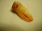
Freshly extracted tooth 34 , exceptionally two roots

Freshly extracted lower molar , exceptionally four roots


Treatment steps of an extraction. Special pliers ( bent over the edge ) were used

Possible complications

- Fracture of the tooth
- Fracture of the root
- Alveolar wall fracture
- Pain, edema (swelling), hematoma (bruise)
- Difficulty opening the mouth up to the jaw clamp
- difficulties swallowing
- dry alveolus : Dry socket (lat .: alveolitis sicca ): clinical picture of wound infection of the jaw bone after a dental removal as a consequence of the disintegration of the blood clot
- Rebleeding
Rare complications
- Opening of the maxillary sinus (in the area of the maxillary posterior teeth)
- A tooth root (or part of it) enters the maxillary sinus
- Damage or loosening of other teeth
- Injury to soft tissue
Very rare complications

- Tear of the maxillary tuberosity (see figure for its position)
- Fracture (break) of the lower jaw or the temporomandibular joint
- Dislocation (dislocation) of the temporomandibular joint
- Ingestion of teeth or parts of teeth
- Aspiration of teeth or parts of teeth

Mouth-antrum connection
The floor of the maxillary sinus is very thin or nonexistent in the area of the root tips of the maxillary posterior teeth. When extracting molars, less often premolars, the maxillary sinus can therefore be opened (which is not a treatment error). After the extraction of such teeth, a so-called nasal blow test ( Valsalva pressure test) is therefore routinely carried out. It is more reliable to probe the alveolus with a button probe for a perforation . If the result is positive, the resulting mouth-antrum connection (mouth-maxillary sinus connection) must be covered with a vestibularly pedicled expansion flap by a surgically experienced dentist , an oral surgeon or an oral surgeon . Otherwise, bacteria and viruses in the oral flora can enter the normally sterile maxillary sinus and cause odontogenic maxillary sinusitis . To do this, the relatively tough periosteum (bone skin) on the inside of the flap is slit so that the mucoperiosteal flap can be stretched sufficiently. The subsequent seam closure must be tight.
As far as possible, the patient should avoid blowing his nose in the first few days after the operation, as the pressure can tear open the closure of the maxillary sinus. Sneezing should be done with your mouth open.
In the case of inflamed maxillary sinuses (e.g. due to the infected root tip), the primary covering with a flap technique often fails because the secretion flowing out (from the nasal cavity via the alveolus into the oral cavity) paves the way for a connection - even if the wound is tightly closed. In this case, the inflammation of the maxillary sinus mucous membrane must be awaited to heal before the plastic covering. If necessary, the maxillary sinus is rinsed over the opened alveolus by an ear, nose and throat specialist.
If tooth fractures occur during extraction, collect all tooth parts to ensure that no tooth parts have been aspirated or left in the wound. Swallowing teeth or tooth parts is associated with only minor risks. A dangerous complication, on the other hand, is aspiration of teeth or tooth parts that require referral to a specialist in ear, nose and throat medicine.
A root dislocated in the maxillary sinus must be removed. This is done by a surgically experienced dentist, a specialist in oral and maxillofacial surgery or a specialist in ear, nose and throat medicine .
Aftercare
Generally, the wound will be checked the following day. If the wound has been sutured, the sutures are removed after about a week, provided that no absorbable suture material that dissolves itself has been used. After a plastic covering of the maxillary sinus, the sutures are removed after ten days. The wound usually closes with primary wound healing during the first few weeks after treatment . The blood clot is the basis for the bony regeneration in the alveolus. The coagulum is slowly organized in connective tissue, with new bone formation starting from the edges after just one week. After about a month, the alveolus is filled with fibrous bone, which is slowly converted into lamellar bone.
If the socket is dry, the wound is revised by curettage of the wound and then inserting a strip of gauze , which is changed daily. In this case, secondary wound healing takes much longer .

Wound of a freshly extracted wisdom tooth, secondary finding: tongue piercing
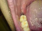
The same wound after two days

The bone compartments (alveoli) of the first lower molar are not yet completely ossified again even after months.


Behavior after the procedure
In the first time after the procedure, correct behavior is necessary for proper wound healing and the avoidance or reduction of complications.
- On the day of the procedure, the wound should be cooled every 20 minutes using an ice pack or cold pack. This prevents swelling. The wound can also be cooled from the inside by sucking ice cubes.
- After a few days, if you are not allergic to chamomile , you can rinse with chamomile to support wound healing after eating.
- Dairy products should be avoided as the lactic acid bacteria they contain could destroy the blood clot that forms.
- The consumption of caffeine (coffee, black tea, energy drinks), nicotine or alcohol should be restricted because these encourage the bleeding tendency.
- The teeth should be carefully cleaned with a soft toothbrush after each food intake.
- The wound area itself should be excluded from cleaning in order to avoid wound healing disorders.
- Physical exertion should be avoided, as the rise in blood pressure can lead to rebleeding.
Follow-up therapy
Is the replacement of the extracted tooth necessary for it are depending on the findings of the various ways dentures , including care provided by an implant available.
Hardship regulation versus social indication for tooth extractions
According to Section 55 SGB V, insured low-wage earners are entitled to partial or full coverage of the treatment costs for standard care, depending on their income and family situation (e.g. life partners increase the income limit).
If another care is desired, the costs of standard care can be deducted from the costs of other care (Section 55, Paragraphs 4 and 5, SGB V).
The hardship rule does not apply if it can be "recognized" that the insured person has not looked after his or her teeth and / or has not made use of the dental checks (Section 55 (1) SGB V).
Use of extracted teeth
More recently, a discussion has developed about the ethical permissibility of using extracted teeth without proof of origin in research and teaching.
Individual evidence
- ^ Gottfried PF Schmuth: Orthodontics , Georg Thieme Verlag, Stuttgart
- ^ Gottfried PF Schmuth: Orthodontics , Georg Thieme Verlag, Stuttgart.
- ^ Research Uni-Leipzig: From the history of tooth extraction - tooth forceps .
- ↑ Johann Jakob Heinrich Bücking: Complete instructions for extracting teeth from Bücking, the knowledge of medicine and the art of wound medicine. Stendal 1782.
- ↑ Dominik Groß : Tooth Extraction. In: Werner E. Gerabek , Bernhard D. Haage, Gundolf Keil , Wolfgang Wegner (eds.): Enzyklopädie Medizingeschichte. De Gruyter, Berlin / New York 2005, ISBN 3-11-015714-4 , p. 1516 f., Here: p. 1516.
- ↑ Ullrich Rainer Otte: Jakob Calmann Linderer (1771-1840). A pioneer in scientific dentistry. Medical dissertation, Würzburg 2002 (with text edition of doctrine of the entire dental operations. 1834, here: pp. 47–55 ( Die Zahnzangen ), 56–64 ( Der Geisfuß ), 64–68 ( The Tooth Key ), 68–73 ( Der Pelikan ) and 73-75 ( Der Ueberwurf ) as well as p. 75 f. ( The lever ) and p. 76 f. ( Pyramid- shaped screw )).
- ↑ Dominik Groß: Tooth Extraction. 2005, p. 1516.
- ↑ a b c d N. Jakse, Extraction customer ( memento from July 10, 2012 in the Internet Archive ), University of Graz.
- ↑ Joachim Gabka / Herbert Harnisch: Operationskurs for dentists , Georg Thieme Verlag, Stuttgart.
- ↑ Joachim Gabka / Herbert Harnisch: Complications and errors in dental treatment , Georg Thieme Verlag, Stuttgart.
- ↑ § 55 SGB V entitlement to benefits. Retrieved July 24, 2018 .
- ↑ Dominik Groß, Christian Lenk, Brigitte Utzig: Normative framework conditions for the recruitment and use of extracted teeth in research and teaching . In: Ethics in Medicine. Volume 28, No. 1, 2016, pp. 21-31.
See also
- Tooth loss
- Wisdom tooth
- Osteotomy
- Evaluation standard of dental services, BEMA numbers 43, 44, 45, 47a and 48
- History of dentistry