
Wisdom tooth
| Classification according to ICD-10 | |
|---|---|
| K00.2 | Abnormalities in the size and shape of the teeth |
| K00.4 | Disturbance of tooth formation |
| K00.6 | Disorders of tooth eruption |
| K00.9 | Disturbance of tooth development, unspecified |
| K01.0 | Retained teeth |
| K01.01 | Impacted teeth |
| K03.3 | Pathological tooth resorption |
| K03.5 | Ankylosis of the teeth |
| ICD-10 online (WHO version 2019) | |


The wisdom tooth ( synonym of third molar , Latin dens sapiens, dens serotinus 'coming late' ) is the eighth tooth in the human dentition when counted from the middle . Normally a person has four wisdom teeth, one in each dental quadrant .
General
Features and name
The change of teeth initially ends with the eruption of the second molar at the age of 12. Wisdom teeth develop later. In some people, mineralization of the wisdom tooth germ is only detectable in the X-ray at the age of 14. They usually only break through in adulthood, sometimes not at all. The name “wisdom tooth” in German is derived from this late breakthrough . The name originally comes from the Persian doctor Avicenna (980-1037) from the Qānūn fī ṭ-Ṭibb (القانون في الطب'Canon of Medicine'), which were translated into Latin as dentes intellectus in the Toledo School of Translators. In Greek they were called Sophronisteres by Kleanthes . In other languages the name is also related to wisdom or understanding, e.g. B. in Japanese ( 知 恵 歯chieba ). However, there are also other linguistic derivations, such as “crouching tooth” in Thai , “youngest tooth” in Indonesian or “unknown to parents” ( 親 知 ら ずoyashirazu ) in Japanese , based on the assumption that the teeth do not appear until the children are out have already moved out of the home. They are called Arabic ضرس العقل / ḍirs al-ʿaql / 'Tooth of the Spirit', in French dent de sagesse , in Italian dente del giudizio .
Shape and appearance
Wisdom teeth deviate from their characteristic anatomical shape more often than other teeth. Wisdom teeth appear with three or five cusps. The number of roots is also different. They can grow together or be bent like a hook, so that extraction of the teeth, if necessary , is difficult. In rare cases, excess wisdom teeth, so-called distomolars, also known as nines, grow behind the wisdom teeth.
Evolutionary development
The considerable differences in the shape and time of the eruption of the wisdom teeth, as well as the occasional complete absence of the tooth systems, is possibly the result of an evolutionary development. It is now widely recognized that the original placenta animals had three incisors , one canine , four premolars and three molars in each half of the jaw . Their tooth formula is therefore 3 · 1 · 4 · 3, their number of teeth was 44. All of the Old World monkeys living today , including chimpanzees and humans, on the other hand, have the tooth formula 2 · 1 · 2 · 3, which means 32 teeth. In modern humans, wisdom teeth can therefore be viewed as a rudiment . The reduction in the number of teeth, which is still ongoing in humans, was - as the fossil Ardi shows - associated with a reduction in the size of the snout and canine teeth several million years ago .
classification


The position of the third molar in relation to the occlusal plane of the second molar and the distance between the mandibular ramus (ascending branch of the lower jaw) and the distal side of the second molar were classified by GJ Pell and BT Gregory. There is also the GB Winter classification, which was modified by WH Archer and GO Kruger.
Classification according to Pell and Gregory
- Class 1. Sufficient space is available between the front edge of the ascending branch and the distal edge of the second molar after the third molar has erupted.
- Class 2. The space available between the anterior margin of the ramus mandibulae and the distal margin of the second molar is smaller than the mesio-distal extension of the crown of the third molar. This means that the distal portion of the crown of the 3rd molar is covered by bones from the ascending branch.
- Class 3. The 3rd molar is completely covered by the bone of the anterior margin of the ascending branch. There is not enough space available.
Health consequences
The imbalance between jaw size and number of teeth leads to the jaw angle, the transition from horizontal to ascending branch of the mandible to the fact that they are frequently not sufficient space more and completely retiniert remain or only partially break through (partial retention). Fully retained teeth usually remain symptom-free, while partially retained teeth often lead to inflammation (see: Dentitio difficilis ), which can develop into abscesses . The cause of such an inflammation is the formation of a hood-shaped gum pocket , which is difficult or impossible to clean. Bacteria can quickly multiply in this pocket with the help of decomposing food particles. As a result, the affected teeth must be removed frequently. Caries can also be favored if wisdom teeth are in contact with a neighboring tooth in such a way that it is difficult to brush there.
In extremely rare cases, the inflammation can turn into a phlegmon , which can be life-threatening.
If the wisdom teeth break through only in one jaw, they lack the antagonist ("opponent"). As a result, they elongate over the occlusal plane until they finally meet the opposing jaw. Wisdom teeth can become "sliding obstacles" that can lead to tooth damage, nocturnal teeth grinding ( bruxism ) and temporomandibular joint problems.
Retained wisdom teeth
“Retiniert” ( Latin retinere re , back, tenere , hold) is the term used to describe a wisdom tooth that has not erupted in accordance with its age. Whether or not a wisdom tooth is retained depends on the age, gender and level of development of the patient. The assessment on the basis of an X-ray is not always clear and requires a follow-up.
The following X-ray images are excerpts from orthopantomograms (OPG):
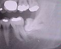
Wisdom tooth 38; retained and displaced; mesial tilt of almost 90 °

Wisdom tooth 38; broken through almost to the occlusal plane; distal wall entirely in bone; fully developed root; Root tips project onto the inferior alveolar nerve

Wisdom tooth 38; retained and displaced - strongly tilted mesially; very short root tips project onto the inferior alveolar nerve

Wisdom teeth 48 and 18; Tooth 48 - no tilting, root only partially formed, crown partially still covered with bones

Wisdom tooth 38 applied; retained - crown wedged under tooth 37; Occlusal crown no longer covered by bone; only one root visible - relatively short but fully developed.

Wisdom tooth 38 applied - almost no root formed; tilted medially.


Wisdom teeth removal
A tooth is removed by loosening the tooth in its tooth socket . For this purpose, the tooth is moved back and forth slowly and with measured force by means of a lever or pliers, whereby the alveolar bone is stretched and the alveolus is expanded. In the case of single-rooted teeth, a rotary movement can also be carried out, whereby the Sharpey fibers , by which the tooth is suspended in the alveolus, tear.
extraction

Many wisdom teeth are regularly in the row of teeth. If necessary, they can be extracted like other teeth . In the lower jaw, in the region of the mandibular angle, the bone is compact and buccally thicker, which means that the alveolus cannot be expanded so easily. In the upper jaw, the bone surrounding the wisdom tooth is cancellous . This makes extraction easier.
Surgical tooth extraction

The osteotomy (surgical removal) of a wisdom tooth is usually carried out under local anesthesia by a surgically experienced dentist , an oral surgeon or an oral surgeon . In the case of very anxious patients, if they wish, analgesic sedation can be carried out in appropriately equipped doctor's offices , which calms the patient down and reduces his or her awareness.
If a particularly difficult tooth extraction is to be expected or if a painless procedure cannot be guaranteed due to conduction or infiltration anesthesia, general anesthesia can also be considered. In cases where all four wisdom teeth are to be removed in one session, general anesthesia is considered.
Surgical removal takes place through an incision on the alveolar ridge, if necessary with a so-called relief incision towards the buccal. After the bone or the tooth has been completely or partially exposed by folding away the gums , the bone covering the tooth is, if necessary, milled away using a bone cutter (Lindemann cutter ) while cooling with an isotonic saline solution . The opening must be large enough for the tooth to fit through.
If the tooth is very firmly anchored in the jaw, the tooth is displaced in the jaw, or the extraction opening is too small, it may be necessary to separate the tooth from the jaw before it is removed. If the wisdom tooth is surgically removed as a tooth germ before it erupts, one speaks of a germectomy . The resulting wound is closed with surgical sutures .
Possible complications


- Pain, edema (swelling), hematoma (bruise)
- Difficulty opening the mouth up to the jaw clamp , difficulty swallowing
- Alveolitis sicca (dry socket , engl. Dry socket ): Clinical picture of wound infection of the jaw bone after a dental removal as a consequence of the decomposition of blood coagulum
- Rebleeding
The following complications are less common:
- Side effects of the anesthetic
- Irritation (irritation) or severing of the inferior alveolar nerve
- Irritation or severing of the lingual nerve
- Fracture (break) of the lower jaw
- Tear off the maxillary tuberosity (see figure for position).
- Opening of the maxillary sinus
- A tooth root (or part of it) enters the maxillary sinus
- Breakage of the injection needle
Behavior after the operation
As a precaution, a pain medication should be prescribed. The pain reliever should not contain acetylsalicylic acid (such as aspirin), as this has a negative effect on blood clotting. Patients with anticoagulant treatment ( Phenprocoumon , Marcumar) can temporarily be switched to low molecular weight heparin preparations , but the current S3 guideline "Dental surgery under oral anticoagulation / platelet aggregation inhibition" sees no advantage here and votes in favor of leaving the original anticoagulant medication. The wound usually closes within the first few weeks after treatment. The treating doctor may prescribe an antibiotic for particularly complicated procedures or if you have had a previous infection .
In the first time after the operation, correct behavior is important for good wound healing and the reduction of the unavoidable sequelae of the operation:
- In the first 24 hours, the wound should be cooled every 20 minutes. This causes less swelling. Cool packs can be used for this.
- Rinsing hinders the natural wound healing process.
- After the extraction / operation, caffeine (coffee, black tea, energy drinks) should be avoided because the increase in blood pressure can cause rebleeding. Alcohol should also be avoided because of its anticoagulant effect. Tobacco smoke increases the risk of wound healing disorders.
- In the first few days after the extraction / operation, milk should be avoided, as the lactic acid bacteria may penetrate the wound when consumed and cause inflammation there.
- On the day of the operation, it is advisable to initially only consume soups and porridge-shaped foods.
- Dental care should be continued as usual if possible. In the first few days, the teeth can be left out in the wound area. But they should be cleaned as soon as possible with a soft brush.
- Alternatively, a mouth rinse containing chlorhexidine can be used. Mouth rinses with at least 0.1% chlorhexidine have an antiseptic effect and reduce the number of germs in the oral cavity. Therefore, these mouthwashes can temporarily replace brushing your teeth and also support the treatment of local inflammations in the oral cavity.
- Sports activities and physical exertion should be avoided as the rising blood pressure can cause secondary bleeding. In addition, strong exertion hinders the body's regeneration and delays wound healing.
- Excessive heat exposure, such as sunbathing, solarium and sauna stays, should be avoided.
literature
- S2k guidelines for surgical removal of wisdom teeth of the German Society for Oral and Maxillofacial Surgery (DGMKG) and the German Society for Dentistry, Oral and Maxillofacial Medicine (DGZMK). In: AWMF online (as of August 2019)
See also
Web links
- Surgical removal of wisdom teeth (PDF; 120 kB) Scientifically proven patient information from the German Dental Association and the German Society for Dentistry, Oral and Maxillofacial Medicine
- Study on pain after wisdom tooth surgery
Individual evidence
- ^ Georg Carabelli von Lunkaszprie: Systematic handbook of dentistry. Volume 1: Historical overview of dentistry. Braumüller and Seidel, Vienna 1831, (digitized version)
- ↑ Winfried Henke , Hartmut Rothe : Stammesgeschichte des Menschen. An introduction. Springer, Berlin a. a. 1999, ISBN 3-540-64831-3 , p. 33 f.
- ↑ Glenn J. Pell, G. Thaddeus Gregory: Report on a ten-year study of a tooth division technique for the removal of impacted teeth. In: American Journal of Orthodontics and Oral Surgery. Volume 28, No. 11, 1942, pp. B660-B666, doi: 10.1016 / S0096-6347 (42) 90021-8 .
- ^ GJ Pell, BT Gregory: Impacted mandibular third molars: classification and modified techniques for removal. In: Dental Digest. Volume 39, 1933, ISSN 0011-8567 , pp. 330-338.
- ↑ George B. Winter: Principles of Exodontia as Applied the Impacted Mandibular Third Molar. A complete Treatise on the Operative Technic with Clinical Diagnoses and Radiographic Interpretations. American Medical Book Co., St. Louis MO 1926.
- ^ W. Harry Archer: Oral and Maxillofacial Surgery. 2 volumes. 5th edition. Saunders, Philadelphia PA 1975, ISBN 0-7216-1362-4 .
- ^ Gustav O. Kruger (Ed.): Oral and Maxillofacial Surgery. 6th edition. Mosby, St. Louis MO 1984, ISBN 0-8016-2793-1 .
- ↑ Impacted Mandibular 3rd Molar Classification. In: Dentistry and Medicine. 2014. Retrieved November 29, 2015.
- ↑ Frank Schneider: Impacted wisdom teeth . (PDF) In: Wisdom Tooth eBook . 2012.
- ↑ Till S Mutzbauer: Pain therapy for wisdom tooth removal . (PDF) In: Pain after wisdom tooth surgery . 2016.
- ↑ awmf.org