
Root canal treatment

In dentistry, root canal treatment is understood as a therapy with the aim of preserving a tooth whose pulp (popularly: "tooth nerve") is vital but irreversibly inflamed or devitalized (dead). The vital or non-vital pulp tissue is removed from the root canal, the root canal is expanded and the infected root dentin surrounding the root canal is removed by filing out. Finally the root canal is filled. Root canal treatment is part of endodontics , which in turn is part of conservative dentistry .
causes

The causes of inflammation of the tooth pulp ( pulpitis ) are varied. Usually there is initially a carious defect that serves as an entry point for bacteria and is painless for a certain period of time. A tooth fracture or treatment trauma , for example due to overheating when grinding the tooth for a tooth crown , can lead to pulpitis, which can be acutely extremely painful. Occasionally, a retrograde inflammation of the pulp can occur if the tooth-holding apparatus is damaged to such an extent that the infection penetrates through the gingival pocket to the tip of the root and from there into the root canal .
In the course of an odontogenic infection , if left untreated, the pulp dies and the germs spread in the system of the root canals. The body reacts with an inflammation of the periodontium ( apical periodontitis ) in the sense of a defense reaction. Apical periodontitis can be acute or chronic . The acute form is often associated with pain and can be difficult to verify radiologically , while chronic apical periodontitis can be seen as a so-called brightening when the bone structure in the area of the root tip breaks down . The brightening appears dark in the X-ray image because the X-ray image is negative .
Indications

Root canal treatment is usually carried out for two indications:
- If the tooth is vital and the pulp is irreversibly damaged, vital extirpation is performed. After local anesthesia , the pulp is removed with an extirpation needle and the system of the root canals is mechanically cleaned using a so-called preparation. The canal wall is filed with files with increasing diameter. After further cleaning processes using sodium hypochlorite solutions , the tooth is closed again using a root canal filling.
- If the tooth is already devitalized, the aim of treatment is to remove the gangrenous pulp and germs from the inside of the tooth. After the tooth has been opened, the system of the root canals is filed and cleaned - as with vital extirpation.
In some cases a vital, healthy tooth has to be devitalized by means of a root canal treatment. This may be necessary in the following cases, if the tooth could otherwise not be restored prosthetically:
- The tooth has an unfavorable, mostly tilted position and should serve as a pillar for a prosthetic restoration, for example a partial denture on telescopic crowns , but due to the degree of tilt it must be ground so far that an opening of the pulp cannot be avoided.
- The vital tooth is fractured at the gum level, the pulp is not opened and the tooth has to be rebuilt with a post . To do this, a fastening pin must be inserted into the root canal.
Aim of treatment
Goals and principles of root canal preparation
- Complete removal of pulp tissue, germs and necrotic material from all canals
- Maintaining the integrity of the periapical tissue or creating the conditions for the healing of existing lesions of endodontic origin
- Preparation exactly up to the endodontic apex
- Uniform processing of the canal walls on all sides without changing the shape of the canal and without excessive weakening of the root
- Shaping to facilitate and optimize the final filling
Goals and principles of root filling
Bacteria-proof and permanent closure of the root canal system with non-resorbable, marginal, radiopaque and biocompatible materials such as gutta-percha . The filling material should also be easily removable again if necessary.
Practical implementation


Access cavity
After applying a rubber dam if necessary, access to the canal system is first created. On the one hand, this must be large enough to be able to carry out the treatment with good visibility, but on the other hand not too large to avoid the unnecessary loss of healthy tooth substance.
cleaning
After local anesthesia , the length of the root canal or canals is determined (using a single x-ray image in connection with special measuring needles or by electrical means through endometry ). The canals are then conically widened (“prepared”) with hand files or machine-driven rotating instruments . By rinsing with different solutions such as For example : NaOCl , 3% H 2 O 2 solution , EDTA or CHX , impurities are removed from the channels, the smear layer is removed and microorganisms are combated. In addition to the commonly used rinsing solutions, a laser can be used to disinfect the canals before filling. The irrigation solutions can be activated by ultrasound to increase the effect. In this way it is also possible to disinfect canal branches and infected dentin areas that are not accessible for instrumental preparation. Dentists specializing in endodontics use a surgical microscope for root canal treatment, which, with its additional light source with coaxial light and magnification, makes it easier to find and view the canal entrances.
Shaping
In order to prepare the canal system in accordance with the filling, it has proven useful to first coronally widen the canal entrances from coronal to apical with Gates-Glidden drills. Further preparation of the canal system can either be carried out with an apical-coronal method such as the step-back technique or, in the case of curved canals, with a coronal-apical method such as the crown-down technique. In addition to removing dentine (for cleaning purposes), the root canal instruments are primarily used to shape the root canal cavities. By processing the root canal walls, the instruments create space to improve the effectiveness of the irrigation solutions and to prepare a defined profile for predictable closure.


filling

The root filling is one of the tooth fillings . After cleaning and shaping the root canals, the cavities are filled. This is mainly done with gutta-percha and a sealing cement, a so-called sealer . The root filling should contain as much gutta-percha as possible and as little sealer as possible, because the gutta-percha is the more biocompatible and stable material.
If an immediate root filling is not possible, a medicated insert is first made. In most cases, a calcium hydroxide preparation (containing iodine) is used, more rarely a cortisone antibiotic preparation. In these cases the root canals are not finally closed until a further treatment session.
The root canal can be filled in the traditional way using cement and a gutta-percha point (one-pin method) or using lateral condensation, where additional gutta-percha points also enable a denser root filling. There is also the option of filling the tooth using thermal root filling techniques. A gutta-percha point is heated and then inserted into the canal, so that it is possible to penetrate the small lumen through the liquefaction of the material in the ramifications of the root canal at the root tip. The sealer material around the root tip can be pressed out here (puff). There is also the option of first inserting a pin and then heating it up in the canal and separating it (so-called downpack) and filling the rest of the canal with liquid gutta-percha (backfill). For the last-mentioned technique, a surgical microscope is advantageous for a better view. In addition, the surgical microscope offers the possibility of finding other smaller canals in the tooth or of covering perforations in the canal with special materials.
research
With the help of nanodiamonds, new types of gutta-percha points are said to successfully combat the growth of bacteria after root canal treatment. These are tiny carbon compounds that can be specifically filled with drugs and thus contribute to an improved effect in the treatment. The gutta-percha points called NDGP are mixed with a proportion of nanodiamonds and the broad spectrum antibiotic amoxicillin . This is intended to prevent residual bacterial infections after root canal treatment.
- composition
Trans-1,4-polyisoprene + ZnO 2 + BaSO 4 + wax
Nanodiamonds + amoxicillin → ND-AMC
example
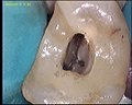
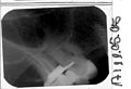
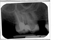
The process of a root canal treatment on tooth 17

X-ray before root canal treatment

Tooth opened, pulp should be removed
Pulp extirpated

Measurement recording

Three channels prepared and shaped

However, upper molars often have four canals

Bottled all four channels
Control recording after root canal treatment
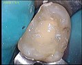
Deck filling
Check-up after 20 months


Possible complications

Under optimal conditions, the chances of success of a root canal treatment are 90 percent. In Germany, however, further treatments are necessary in 60 to 50 percent of cases, as ideal conditions are seldom given and complications often occur. Root canal treatment complications can be caused by:
- inaccessible canal sections (obstruction of the canal lumen by denticles , strong curvature of the root, ramifications),
- broken instruments,
- particularly stubborn microorganisms such as Enterococcus faecalis or Candida albicans , which penetrate up to 0.4 mm deep into dentinal tubules and can survive as a monoinfection,
- additional periodontal damage to the tooth,
- Via falsa ("wrong way") - iatrogenic (caused by the doctor) perforation of the root or
- Fractures of the root.
In some cases, an apicectomy with retrograde root filling is indicated. Alternatively, a revision of the root canal treatment may be appropriate, which should generally be given preference over a tip resection.
- see in more detail: Alternatives to apical resection
Aspects of health insurance law
In Germany, the health insurance guidelines for root canal treatment were changed on January 1, 2004. A root canal treatment can only be provided by the dentist at the expense of the health insurance if the guidelines of the Federal Joint Committee (G-BA) are met for the respective tooth . Endodontic treatment of teeth that do not comply with these guidelines can be arranged privately with the statutory insured patient according to the fee schedule for dentists (GOZ).
Root canal treatment on molars is usually indicated, though
- so that a closed row of teeth can be obtained,
- a one-sided free-end situation is avoided,
- This is the only way to preserve functional dentures.
In addition, the guidelines of the G-BA further limit the contract dentist's root canal treatment.
9.1 Treatment within the framework of contract dental care is only indicated if it is possible to process and fill the root canal up to or close to the tip of the root.
9.4 In the case of pulp-dead teeth with a pathological change at the root tip diagnosed in the X-ray image, a critical check must be made in the prognosis as to whether the attempt to preserve the tooth through conservative or conservative-surgical treatment is being undertaken. For the therapy of teeth with root canal fillings and apical changes, surgical measures are primarily indicated.
9.5 In the case of combined periodontal and endodontic lesions, the preservation of the teeth must be critically examined with regard to the periodontal and endodontic prognosis.
10. As a rule, the removal of a tooth is indicated if it cannot be preserved according to the criteria described in these guidelines. A tooth that is not worth preserving according to these guidelines should be removed. Any other treatment of teeth that are not worth preserving is not part of the contract dental care.
According to Section 8 (7) BMV-Z, additional payments for contractual services are not permitted. However, services that are not included in the catalog of statutory health insurance and are related to root canal treatment may be offered to the patient outside the contract and additionally agreed without the entire treatment having to be billed privately. A decision about the need for additional services is made by the dentist and usually agreed in writing between him and the patient prior to the treatment based on the GOZ. A later assumption of these optional benefits is not covered by German statutory health insurance, but some private supplementary insurance can partially reimburse them.
Extraction of stem cells from the pulp of the milk teeth
The deciduous teeth are suitable as a source of stem cells . The cells in the pulp can be extracted, cultivated with a special growth agent and finally preserved for medical purposes. The stem cells can be used in dentistry for the regeneration of the dental pulp in adults. With the help of transplanting stem cells as part of tissue engineering , parts of the root canals can be renewed.
See also
- Vertucci classification
- Number of roots and root canals
- Evaluation standard of dental services, BEMA numbers 28, 32 and 35
Web links
- Scientific statements of the German Society for Dentistry, Oral and Maxillofacial Medicine, including several on conservative dentistry and endodontic topics
- Technology report on root canal treatment on molars (incl. Success rates) (PDF; 607 kB)
- German Society for Endodontics
- Austrian Society for Endodontics
- Root canal treatment
Individual evidence
- ↑ modified from Michael Hülsmann: Endodontie . In: Ott, RW, H.-P. Vollmer, WE Krug (ed.): Clinic and practice guide dentistry . Georg Thieme Verlag, Stuttgart, New York 2003, ISBN 3-13-131781-7 , pp. 194 .
- ↑ Michael Hülsmann: Endodontics . In: Ott, RW, H.-P. Vollmer, WE Krug (ed.): Clinic and practice guide dentistry . Georg Thieme Verlag, Stuttgart, New York 2003, ISBN 3-13-131781-7 , pp. 201 f .
- ↑ Good clinical practice: The root canal treatment (PDF) Joint statement of the DGZ and the DGZMK, as of July 23, 2007, V 1.b Source: DZZ 60 (2005) 8. Accessed on July 14, 2016.
- ^ Peter HA Guldener, Kaare Langeland: Endodontologie , Thieme Verlag, Stuttgart, New York 1982, ISBN 3-13-610001-8 , pp. 203 ff.
- ^ Peter HA Guldener, Kaare Langeland: Endodontologie , Thieme Verlag, Stuttgart, New York 1982, ISBN 3-13-610001-8 , p. 266.
- ↑ Michael Hülsmann: Endodontics . In: Ott, RW, H.-P. Vollmer, WE Krug (ed.): Clinic and practice guide dentistry . Georg Thieme Verlag, Stuttgart / New York 2003, ISBN 3-13-131781-7 , p. 200 f .
- ↑ Dong-Keun Lee, Sue Vin Kim et al. a .: Nanodiamond - Gutta Percha Composite Biomaterials for Root Canal Therapy. In: ACS Nano. 2015, p. 151021080042004, doi: 10.1021 / acsnano.5b05718 .
- ↑ Nano Diamonds might prevent tooth loss after root canals , UCLA School of Dentistry. Retrieved October 30, 2015.
- ↑ Endodontics online - An initiative to promote endodontics, accessed on April 4, 2016
- ↑ Michael Hülsmann: Endodontics . In: Ott, RW, H.-P. Vollmer, WE Krug (ed.): Clinic and practice guide dentistry . Georg Thieme Verlag, Stuttgart, New York 2003, ISBN 3-13-131781-7 , pp. 205 .
- ↑ Guidelines of the Federal Joint Committee for Sufficient, Appropriate and Economical Contract Dental Care from March 1, 2006 (PDF)
- ↑ BEMA guidelines (excerpt; PDF; 480 kB)
- ↑ THE Commentary BEMA and GOZ - "The Commentary BEMA and GOZ" is the standard work for dental accounting. Accessed on September 23, 2018 (access to the paid version can be guaranteed for 10 days via a free test account).
- ↑ Suseela Keerti Popuri: Concerns of a Pediatric Dentist in Dental Stem Cells: An Overview . Ed .: The open dentistry journal. 2018, p. 596-604 .
- ↑ J. Jobst: Extraction of stem cells from milk teeth. In: Kigorosa. Roman Safreider, January 14, 2019, accessed on March 11, 2019 .
- ↑ G. Schmalz: On the way to the new pulp: can we regenerate the pulp? Ed .: Austrian Dental Congress 2012 and Symposium for Pediatric Dentistry. Volume 109.Springer Vienna, September 2012, p. 52-96 .