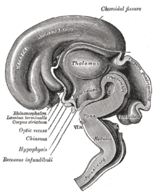
Thalamus
The thalamus (from Greek. Θάλαμος thálamos "bed chamber", "chamber") forms the largest part of the diencephalon . It is made up of many core areas that have a particularly strong connection to the entire cerebral cortex .
In the course of brain development, the thalamus splits into two parts. The “actual” thalamus must therefore be more precisely referred to as the dorsal thalamus . Its task is to modulate the incoming and outgoing information to the cerebrum and thus the cortical excitation. The thalamus ventralis (not to be confused with the ventral core group of the thalamus dorsalis) controls and modulates the excitation of the actual thalamus (dorsalis). Alternatively, the term pair thalamus (for thalamus dorsalis) and subthalamus (for thalamus ventralis) is used.
In most people, the right and left thalamus are developed together over a thin connective tissue bridge , the Adhaesio interthalamica (also massa intermedia ). However, this only contains crossing fibers ( commissures ) in exceptional cases .
Thalamic nuclei
Specific and unspecific thalamic nuclei are distinguished according to their efferents and, above all, their influence on the cerebral cortex. However, this distinction is controversial because the separation is not as sharp as was assumed: unspecific nuclei sometimes have narrowly delimited projections and the efferents of specific thalamic nuclei are often not limited to one cortex area. For the sake of clarity, however, the subdivision is often made.
Specific thalamic nuclei
The specific thalamic nuclei are collectively referred to as the palliothalamus . They are each connected to definable areas of the cerebral cortex and are therefore referred to as specific. You receive sensory (touch, vibration, pain) and sensory (sight, hearing, taste) impulses from the periphery (sensory organs) and, after switching over, forward them to the responsible areas in the cerebral cortex via the thalamic radiation . In addition, information from motor centers ( cerebellum , basal ganglia ) is transmitted to the motor areas of the cerebral cortex.
The specific thalamic nuclei are:
- anterior nucleus group ( nuclei anteriores )
- medial core group ( Nuclei mediales )
- ventral nucleus group ( nuclei ventrales )
-
Nucleus ventralis anterolateralis
- Nucleus ventralis anterior (VA)
- Nucleus ventralis lateralis (VL)
- Nucleus ventralis intermedius (VIM)
- Nucleus ventralis posterior (VP)
-
Nucleus ventralis anterolateralis
- posterior nucleus group ( nuclei posteriores )
- dorsal nucleus group ( Nuclei dorsales )
- Metathalamus , consisting of:
Nonspecific thalamic nuclei
The unspecific thalamic nuclei , also known as the truncothalamus , have only a few direct connections to the cerebral cortex and, in contrast to those of the specific thalamic nuclei, they cannot be narrowed down to specific areas. They are efferent mainly connected to the specific thalamic nuclei, so they control them. This also leads to a non-specific activation of the cerebral cortex when the unspecific thalamic nuclei are excited. They receive afferents primarily from the reticular formation , from the cerebellum and from the basal ganglia.
Important non-specific cores:
function
Supplying (afferent) nerve cells carry information from the body and the sensory organs into the thalamus, where they are switched in the “specific thalamic nuclei” to a subsequent nerve cell that leads to the cerebral cortex . This switching ( synapse ) enables primitive information processing, in which the thalamus acts as a filter and decides which information is so important for the organism at the moment that it should be passed on to the cerebral cortex and made aware. The thalamus is therefore often referred to as the “gateway to consciousness”. This switching / information processing is controlled by the "unspecific thalamic nuclei", which in turn receive their input from other areas of the brain. This regulation is necessary so that the thalamus can adapt decisions (“What is currently important?”) To the overall situation (e.g. sleep, foraging, mating time). The nerve cells supplying the thalamus are mostly crossed over, so that each side of the thalamus represents the opposite half of the body .
Opioid receptors are also located in the thalamus .
Ventral thalamus
For the principle of the interconnection of the thalamus (dorsalis) it is crucial that all corticothalamic and thalamocortical projections are reciprocal. This means that an excitation of the cortex by the thalamus (dorsalis) now also reciprocally results in an excitation of the thalamus (dorsalis) by the cortex . This also applies to inhibitory impulses from the thalamus (dorsalis). It quickly becomes clear that reciprocal excitation would quickly lead to cortical overexcitation, while inhibition of the cortex via reciprocal inhibition of the thalamus (dorsalis) would mean that the cortex could no longer be activated at rest.
Therefore, there must be a second regulatory station for the thalamus (dorsalis). This is the thalamus ventralis (also subthalamus ), which is split off from the thalamus (dorsalis) in evolutionary terms. The thalamus ventralis includes important core areas that are functionally assigned to the basal ganglia ( nucleus subthalamicus and globus pallidus ). Due to a complicated interconnection within the basal ganglia, these nuclei control the thalamus (dorsalis) directly (in the case of the internal segment of the globus pallidus) or indirectly. See the article on the function of the basal ganglia .
Another important control instance of the thalamus ventralis is the nucleus reticularis thalami ( reticulated thalamic nucleus ), which surrounds the thalamus (dorsalis) in a reticulated manner. It is structured internally in such a way that there are corresponding areas in the reticular nucleus for each area of the thalamus (dorsalis). The reticular nucleus also receives fibers of the cortex, which receive copies of all projections of the cortex onto the thalamus (dorsalis) and, with a time delay, now project antagonistically onto the corresponding thalamic area. An exciting projection onto the thalamus (dorsalis) thus effects a delayed inhibition of the excited thalamic nucleus via the reticular nucleus. An inhibition of the thalamus (dorsalis) is canceled analogously via the nucleus reticularis. This solves the “dilemma” outlined above.
Diseases
Damage to the thalamus mainly affects the opposite (contralateral) side of the body and leads to the following types of disorders:
- Ataxia
- Hemianopia
- Hemiparesis
- Loss of sensitivity
- Thalamic pain
literature
- E. Eitschberger: Development and chemodifferentiation of the rat thalamus. Springer Verlag, Berlin / Heidelberg 1970.
- Joachim K. Krauss, Jens Volkmann: Deep brain stimulation. Springer Nature Switzerland AG 2019.
- Justin L. Song: Thalamus. Anatomy - Functions and Disorders, Nova Science Publishers 2011, ISBN 978-1-6132-4152-3 .
- R. Nieuwenhuys, J. Voogd, C. van Huijzen: The central nervous system of humans. An atlas with accompanying text, Springer Verlag, Berlin / Heidelberg 1980, ISBN 978-3-540-10031-7 .
See also
Web links
- Article . In: Scholarpedia . (English, including references)
- The thalamus: gateway to consciousness and rhythm generator in the brain (accessed February 20, 2020)
- Evolution of Nervous Systems: II Vertebrates (accessed February 20, 2020)
- LEXICON OF BIOLOGY: Thalamus (accessed February 20, 2020)
- Characterization of glial cells in the mouse thalamus (accessed February 20, 2020)


