Metathalamus
As metathalamus ( gr. Μετά meta 'above, according behind'; 'Nachthalamus') is a part of the brain of mammals , more specifically of the diencephalon ( diencephalon hereinafter), namely a portion of the thalamus to the dorsal (posterior) portion. The metathalamus consists of two pairs of lateral and central knee cusps ( corpora geniculata laterale et mediale ).
Corpus geniculatum laterale
The lateral knee hump ( Corpus geniculatum laterale , CGL ) is a core area in which about 90% of the axons of the visual pathway ( Tractus opticus ) end. However, the neurons of the CGL receive input not only from the retina, but also from other thalamic nuclei and from neurons of the CGL itself, as well as from the cerebral cortex and the upper mounds of the midbrain . All this information is integrated, modified and, to a large extent, passed on to the visual cortex via the visual radiation ( Radiatio optica ) . In addition, there are efferents to nuclear areas in the superior colliculus , to the pretectal nuclei and to the suprachiasmatic nucleus of the hypothalamus .
(The remaining 10% of the optic nerve fibers end directly at nuclei in the superior colliculi, in the area praetectalis or in the hypothalamus. Their information is used, among other things, for the reflex head and eye movements , the pupillary reflex and the day-night rhythm .)
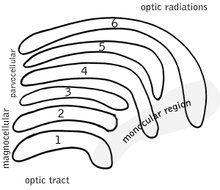
The core areas of the corpus geniculatum laterale show on each side a structure of six cap-like curved, approximately concentrically arranged layers, for which a large cell (magnocellular) area ( nucleus ventralis corporis geniculati lateralis ) and a small cell (parvocellular) area ( nucleus dorsalis corporis geniculati lateralis) ) distinguish.
Division into layers
Assignment of the half of the field of view
The CGL is made up of six layers, which are referred to as layers 1 to 6 from ventral to dorsal. Slices 1, 4 and 6 receive input from the contralateral eye, slices 2, 3 and 5 from the ipsilateral eye, in each case via the half of the visual field opposite the CGL . The right CGL therefore receives information about the left half of the visual field, which comes from the left eye (layers 1, 4 and 6), and information about the left half of the visual field from the right eye (layers 2, 3 and 5). Thus, each half of the brain receives information from both eyes, but initially only via one mutual half of the visual field (see also: contralaterality of the forebrain ). This separation is retained in the projections of the CGL to the primary visual cortex (V1, area striata, area 17) and thus reaches neocortical layer 4 of the visual cortex. Only in the further cortical processing is the information about both halves of the field of view then processed together to different degrees.
The two ventral layers 1 and 2 of the CGL are called the magnocellular layers (or also the ventral nucleus), the layers 3 to 6 as the parvocellular layers (or also the dorsal nucleus). You will receive input from different types of retinal ganglion cells . Between the six layers are the coniocellular layers of very small cells, the function of which is not yet fully understood. You will receive input from retinal ganglion cells of the bistratified type.
Magnocellular layers (nucleus ventralis CGL)
The neurons of the two ventral layers of the CGL are characterized by larger cell bodies, more extensive dendrite trees and stronger axons compared to the neurons of the other layers. The larger diameter of their axons results in a very high conduction speed to downstream units. The input cells of this layer - the magnocellular retinal ganglion cells of the parasol type - are characterized by a large receptive field. However, they are completely insensitive to color perception, since their receptive fields in both the center and the periphery consist of the same type of cone ( color perception is only possible if the center and periphery of a receptive field receive input from different types of cones). The function of the magnocellular system has been examined in particular with lesion studies in monkeys: The magno- or parvocellular layers were selectively deactivated by pharmaceutical methods. Monkeys, in which the magnocellular layers of the CGL were switched off, showed clear deficits in the perception of movement, location and speed. However, color vision and visual accuracy were not affected. Therefore one sees in the magnocellular layers an essential input for the "dorsal route" of the visual processing in the brain, which v. a. Movement, place and action perception processed. Starting from V1, the dorsal route comprises different areas in the direction of the parietal cortex, such as Area MT, MST, MIT.
Parvocellular layers (nucleus dorsalis CGL)
In the third to sixth layers of the CGL there are parvocellular neurons with smaller cell bodies, more restricted dendrite trees and thinner axons (conditionally slower conduction speed). You receive input from parvocellular retinal ganglion cells of the midget type, which have a smaller receptive field and thus enable a higher visual resolution. Color perception is only possible with the parvocellular system. The center of a receptive field of a parvocellular ganglion cell consists of a different type of cone than its periphery. A color can be perceived by comparing the excitation strength of the types of cones. There are three types of cones, each of which shows the greatest sensitivity for short, medium or long-wave light (blue, green or red).
The parvocellular system is necessary for a perception with visual resolution of finer accuracy (size, shape, texture), as well as for color perception. It is considered to be the most important input for the "ventral route" of visual processing in the brain, which starts from V1 in the ventral direction to the inferior temporal lobe (e.g. V4, fusiform gyrus). It is responsible for shape, sharpness, color perception and object recognition.
Corpus geniculatum mediale
The medial knee hump ( corpus geniculatum mediale ) is the switching station of the auditory pathway . Their information reaches the knee cusp via the lemniscus lateralis and colliculus inferior and from there via the radio acustica to the auditory cortex in the temporal lobe . The corpus geniculatum mediale is anatomically and functionally divided into three parts.
Pars ventralis
The ventral part is the main part of the auditory thalamus. It encompasses the entire specific ( lemniscale ) component of the auditory pathway, i.e. the main strand of acoustic information processing. It receives ascending fibers from the central core of the inferior colliculus on the same side and pulls with outgoing fibers to the primary auditory cortex (AI) and anterior (anterior) auditory field (AAF) on the same side. In the course of a feedback, it receives descending fibers from all areas of the cortex that are involved in the hearing system.
The neurons and the catchment area of their dendrites are arranged in layers ( laminae ). Several neighboring neuron layers form a functional layer network. In the area of a composite there is a fine scaling according to acoustic frequencies, while from composite to composite there is a coarse scaling at an octave interval .
The function of the pars ventralis consists in the combination of any kind of frequency-specific information and its modulation by descending input from the cortex before it is sent to the cortex for further processing. A special effect of anatomy and function is possibly the octave identity known in music .
Pars dorsalis
The pars dorsalis is part of the non-specific (extralemniscal) component of the auditory pathway, which combines auditory and non-auditory information. It receives ascending fibers from the dorsal cortex of the equilateral inferior colliculus and from the somatosensory system. It pulls with outgoing fibers to the secondary and tertiary auditory cortex on the same side (AII and others). In the course of a feedback, it receives descending fibers from all areas of the cortex that are involved in the hearing system.
The function of the pars dorsalis consists in the combination of any kind of non-frequency-specific auditory and non-auditory information. Again, this process is subject to modulation by descending input from the cortex.
Pars medialis
The pars medialis is also part of the non-specific (extralemniscale) component of the auditory pathway. It receives ascending fibers from all three main departments of the equilateral inferior colliculus , from the superior olive (nucleus olivaris superior), from the superior colliculus and from the equilibrium system . With outgoing fibers, it pulls to all areas of the auditory cortex (AI, AII, etc.) and also to the amygdala , a core area of the limbic system . In the course of a feedback, it receives descending fibers from all areas of the cortex that are involved in the hearing system.
The function of the pars medialis consists in the combination of any kind of non-frequency-specific auditory and non-auditory information. Through the connection with the amygdala, this core area is involved in the acoustic conditioning of feelings. Furthermore, like the other main parts of the corpus geniculatum mediale , it is subject to modulation by descending input from the cortex.
Individual evidence
- ↑ JS Cetas, RO Price, DS Velenovsky, DG Sinex, NT McMullen: Frequency organization and cellular lamination in the medial geniculate body of the rabbit. In: Hearing research. Volume 155, Numbers 1-2, May 2001, ISSN 0378-5955 , pp. 113-123, PMID 11335081 .
- ↑ James O. Pickles: Auditory pathways: anatomy and physiology . In: Gastone G. Celesia, Gregory Hickok (Eds.): The Human Auditory System: Fundamental Organization and Clinical Disorders. Volume 129 of Handbook of Clinical Neurology , Burlington: Elsevier Science 2015, 722 pp, pp. 3-25, ISBN 0444626298 , pp. 13-15.


