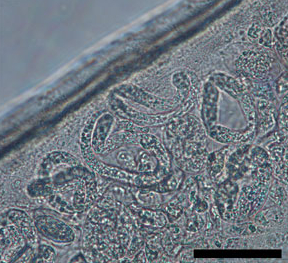
Dirofilaria repens
| Dirofilaria repens | ||||||||||||
|---|---|---|---|---|---|---|---|---|---|---|---|---|

Microfilariae of D. repens in the uterus of a female worm (line: 50 μm) |
||||||||||||
| Systematics | ||||||||||||
|
||||||||||||
| Scientific name | ||||||||||||
| Dirofilaria repens | ||||||||||||
| Railliet & Henry , 1911 |
The nematode Dirofilaria repens is a parasite of the subcutaneous tissue of dogs . Mosquitoes act as intermediate hosts and carriers . The main distribution area of the parasite is southern Europe, but increasingly northern parts are being populated. It causes cutaneous dirofilariasis , one of the nematode infections in dogs ; human infestation is also possible.
morphology
Adult females are 13-17 cm long and 460-650 μm wide, while males are shorter, they are 5-7 cm long. L1 larvae shed from the females are 330-370 µm long and 6-8 µm wide. Adult worms show typical raised longitudinal stripes in the cuticle , the top layer of skin. The strips are spaced 4–24 μm apart.
In the female animal, the vulva is about 1800 μm from the head end. The anus is about 100 μm in front of the tail end. Both sexes have inconspicuous papillae. At the tail end of the male there are two small tail wings, the preanal and postanal papilla, as well as two asymmetrical spicula, the mating organs.
Life cycle
Mosquitoes ingest infectious larvae (microfilariae) with the blood of infected hosts. In the mosquito they develop into third larvae. The duration of this development process depends on the temperature and can range from 8–10 days at 28–30 ° C, 11–12 days at 24 ° C and 16–20 days at 22 ° C. When sucking, third larvae are transferred to the new host. There they develop into fully grown worms that colonize the subcutaneous tissue, mate and in turn form microfilariae again, which can be detected in the blood of infected hosts. The prepatency is comparatively long at 27–34 weeks. Dirofilaria repens can linger in the host's body for up to 7 years.
Hosts and vectors
The main hosts of Dirofilaria repens are domestic dogs, in their bodies the parasites develop into sexually mature animals, mate and produce larvae (microfilariae). Microfilariae can only rarely be detected in the blood of infected cats and wild carnivora such as foxes. In humans, the worms do not normally mate and consequently do not multiply.
Numerous mosquito species have been identified as vectors (carriers and intermediate hosts).
Clinical picture
The infestation with D. repens occasionally causes skin lumps, swellings, itching, abscesses and hair loss, but often runs completely without clinical symptoms. The acid phosphatase reaction can be used for diagnosis . So far, one single case has become known where an infection with D. repens led to meningoencephalitis in a 45-year-old man . On November 9, 2013, Spiegel Online reported a case where one eye was affected.
Extension of the endemic areas in Europe to the north
Dirofilaria repens occurs mainly in southern, southern, eastern and western Europe and in large parts of Asia and Africa. In Greece, infestation rates between 7 and 22% were determined in domestic dogs, in Sicily 2.3% and in France 1.3%. The parasite is not native to the USA, Japan and Australia.
Up to the end of the twentieth century Dirofilaria repens was native to Europe mainly in the Mediterranean area and cases of illness in more northern regions were a result of trips to these areas. However, in the first decade thereafter, there were increasing reports of locally acquired infections in northeastern Europe. In addition to the humidity, the temperature is an essential factor that influences the development of the larvae in the mosquito. Climatic changes are responsible for the fact that the conditions for the development of the larvae of Dirofilaria repens in mosquitoes have been given in Brandenburg since 2001 (until the publication with the report was written in 2012) . Another important factor in the spread are infected dogs brought from endemic areas to more northerly countries and dogs that become infected while traveling to endemic areas. They ensure that mosquitoes can ingest larvae and spread the parasite. Humans can serve as a false host for Dirofilaria repens . While the parasite had not previously been detected in native mosquito species, the Bernhard Nocht Institute for Tropical Medicine reported in 2013 that this filariae was found in several mosquito traps in the Eberswalde area in Brandenburg in 2011 and 2012. In 2014, a case of cutaneous dirofilariasis was detected for the first time in a man in Saxony-Anhalt who had never been in southern Europe. These findings suggest that the filariae, which was previously native to southern Europe, has meanwhile also become native to some areas of Central Europe.
literature
- Claudio Genchi, Marco Genchi, Gabriele Petry, Eva Maria Kruedewagen, Roland Schaper: Evaluation of the Efficacy of Imidacloprid 10% / Moxidectin 2.5% (Advocate®, Advantage® Multi, Bayer) for the Prevention of Dirofilaria repens Infection in Dogs. In: Parasitology Research. 112, 2013, pp. 81–89, doi: 10.1007 / s00436-013-3283-9 (pdf, 5.1 MB; article with a photo of a male Dirofilaria repens ).
- P. Jokelainen, PF Mõtsküla, P. Heikkinen, E. Ülevaino, A. Oksanen, B. Lassen: Dirofilaria repens Microfilaremia in Three Dogs in Estonia. In: Vector borne and zoonotic diseases. Volume 16, number 2, February 2016, pp. 136-138, doi: 10.1089 / vbz.2015.1833 , PMID 26789635 .
Web links
- Dirofilaria repens at the NCBI
- Photos by Dirofilaria repens in the Open-i Project. There are also electron microscopic images that reveal morphological properties.
- YouTube: Surgical removal on a dog
Individual evidence
- ↑ Centers for Disease Control & Prevention, Center for Global Health, cdc.gov: Filariasis (February 13, 2018)
- ↑ a b Claudio Genchi, Marco Genchi, Gabriele Petry, Eva Maria Kruedewagen, Roland Schaper: Evaluation of the Efficacy of Imidacloprid 10% / Moxidectin 2.5% (Advocate®, Advantage® Multi, Bayer) for the Prevention of Dirofilaria repens Infection in Dogs . In: Parasitology Research. 112, 2013, pp. 81-89, doi: 10.1007 / s00436-013-3283-9 .
- ↑ a b M. W. Service, RW Ashford u. a .: Encyclopedia of arthropod-transmitted infections of man and domesticated animals Wallingford, Oxon, UK; New York, NY, USA: CABI Pub., 2001, ISBN 0-85199-473-3 , p. 145 ( limited preview in Google Book Search).
- ↑ Eva Bocková include: Dirofilaria repens microfilariae in Aedes vexans mosquitoes in Slovakia . In: Parasitology Research . tape 112 , no. 10 , October 2013, p. 3465-3470 , doi : 10.1007 / s00436-013-3526-9 .
- ↑ a b R. Sassnau, C. Genchi: Qualitative risk assessment for the endemization of Dirofilaria repens in the state of Brandenburg (Germany) based on temperature-dependent vector competence. In: Parasitology Research. 112, 2013, pp. 2647-2652, doi: 10.1007 / s00436-013-3431-2 .
- ↑ HA Melsom, YES Kurtz neck, K. Qvortrup, R. Bargum, TS Barfod, M. la Cour, S. Heegaard: Subconjunctival Dirofilaria repens infestation: A Light and Scanning Electron Microscopy Study. In: The open ophthalmology journal. Volume 5, 2011, ISSN 1874-3641 , pp. 21-24, doi: 10.2174 / 1874364101105010021 , PMID 21738560 , PMC 3104560 (free full text).
- ↑ L. Keller et al: case report and review of the literature on cutaneous dirofilariasis. In: Veterinary practice small animals . 35 2007, pp. 31-34.
- ^ S. Poppert, M. Hodapp, A. Krueger, G. Hegasy, WD Niesen, WV Kern, E. Tannich: Dirofilaria repens infection and concomitant meningoencephalitis. In: Emerging infectious diseases. Volume 15, Number 11, 2009, ISSN 1080-6059 , pp. 1844-1846. doi: 10.3201 / eid1511.090936 . PMID 19891881 , PMC 2857255 (free full text).
- ↑ A puzzling patient: What caught the eye? accessed on September 20, 2017
- ↑ Ramin Khoramnia, MD, Aharon Wegner, MD: Subconjunctival Dirofilaria repens In: The new england journal of medicine
- ↑ Josef Boch among others: Cutaneous Dirofilariosis. In: Thomas Schnieder (Ed.): Veterinary Parasitology. Paul Parey, 2006, ISBN 3-8304-4135-5 , p. 511.
- ↑ Dog skin worm Dirofilaria repens first detected in German mosquitoes. (PDF; 486 kB) Bernhard Nocht Institute for Tropical Medicine, accessed on August 31, 2013 (press release No. 01/2013).
- ↑ D. Tappe et al .: A case of autochthonous human Dirofilaria infection, Germany. In: Eurosurveillance March 2014.
