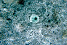
Entamoeba histolytica
| Entamoeba histolytica | ||||||||||||
|---|---|---|---|---|---|---|---|---|---|---|---|---|
|
Entamoeba histolytica ( Magna -form, |
||||||||||||
| Systematics | ||||||||||||
|
||||||||||||
| Scientific name | ||||||||||||
| Entamoeba histolytica | ||||||||||||
| Schaudinn , 1903 |
Entamoeba histolytica is a unicellular parasite that belongs to the Entamoebidae . It is the cause of amoebic dysentery , mainly affects humans and, under experimental conditions, other mammals as well .
features
Entamoeba histolytica is a unicellular eukaryotic organism without mitochondria , lives anaerobically , has a heterotrophic diet and is capable of phagocytosing other cells such as bacteria . Because of its changeable body shape, combined with locomotion via pseudopodia (pseudopodia), it is - like various other species - called an amoeba . This amoeboid protist belongs to the genus Entamoeba . E. histolytica goes through two stages in its life cycle: that of an immobile cyst and that of a trophozoite , which mainly colonizes the human intestinal tract. Its Minuta form is 10 to 15 micrometers in size, the Magna form 25 to 40 micrometers. The cysts that form are tetraploid ; when mature they contain four cell nuclei and reach a size of around 20 micrometers.
Life cycle
These organisms mostly reach the small intestine of the host organism via the upper digestive tract as cysts with contaminated liquid or solid food. Here, a quadricuclear cell stage emerges from the cyst envelope, from which eight small mononuclear trophozoites of E. histolytica emerge through multiple division and colonize the large intestine. In most cases, these form a commensal with the host . The trophozoites can multiply through binary cell division. They can also form cysts that leave the intestines with the faeces . With massive infestation, the amount excreted can easily amount to 100 million cysts per day.
Harmful effect
In 1875, the Russian doctor Fedor Lösch in St. Petersburg described the massive occurrence of amoeba in dysentery for the first time and called it amoeba coli . The pathogen was given the name Entamoeba histolytica in 1903 from the German zoologist Fritz Schaudinn , who researched this protozoon more closely - and contracted an infection in the process.

Infections with Entamoeba histolytica are known as amebiasis . They run in 80–90% of cases without any symptoms. However, asymptomatic infected people contribute to the spread of the disease via cysts excreted in the stool - which can remain infectious for several weeks. The main host, along with some other primate species, is humans. Cysts are only formed from the forms that live in the intestine.
Signs of illness develop in 10–20% of those infected. The colonizing trophozoites can be divided into two forms, the small Minuta and the larger Magna form. Minuta forms resemble other, non-pathogenic Entamoebia species and live in the large intestinal lumen of the host on intact mucosa; this type of colonization usually causes no symptoms.
Diarrhea with abdominal pain are clear signs of irritation of the intestinal epithelium due to the pathogenic form. The Magna form is able to actively penetrate the colon tissue and dissolve it using special enzymes. It can also phagocytize erythrocytes . Such invasive amebiasis can - intestinally - lead to inflammation ( colitis ) and tissue defects ( ulcerations ) in the intestinal tract . If trophozoites get into the bloodstream, they can - extraintestinally - settle in the liver and other organs and cause abscesses there . Liver abscesses are by far the most common form of extraintestinal amebiasis. Three quarters of these cases are not preceded by acute amoebic dysentery.
The clinical picture with diarrhea caused by Entamoeba histolytica is also called dysentery , more precisely amoebic dysentery . Invasive amebiasis , which can manifest both intestinally and extraintestinally , is used to refer to invasive amebiasis in the host's body tissue . This invasion of E. histolytica is facilitated by the release of tissue- dissolving ("histolytic") secretions; purulent ulcers develop in connection with the host's own defensive reactions ; they can break open, but - unlike with bacterial ones - puncture is only advisable in exceptional cases.
In addition to abdominal pain and jelly-like diarrhea , other symptoms can occur. Serious complications are peritonitis and abscesses; they usually heal completely with adequate drug therapy. If left untreated, amoebic dysentery can lead to death due to the loss of fluid through dehydration ( desiccosis ). It is not uncommon for the disease to break out years or decades after the infection .
distribution
Entamoeba histolytica is widespread around the world and is particularly found in areas with poor hygienic conditions, in sewage or in polluted drinking water. Outbreaks are recorded following disasters when there is insufficiently pure drinking water available. Faults in the sewage system can also cause amoebic dysentery, for example over 1,000 cases with 58 deaths were observed at the 1933 World Exhibition in Chicago , caused by the leakage of sewage into the drinking water supply.
Entamoeba dispar is more widespread , a species that was previously assigned to Entamoeba histolytica . It does not develop any magna forms and is visually indistinguishable from the minuta form of Entamoeba histolytica . It usually only causes diarrhea that goes away on its own.
Individual evidence
- ↑ a b L. Ben Ayed, S. Sabbahi: Entamoeba histolytica. In: Global Water Pathogen Project (UNESCO). Part 3 Protists, October 2017, doi: 10.14321 / waterpathogens.34 .
- ↑ J. Blessmann, IK Ali, PA Nu, BT Dinh, TQ Viet, AL Van, CG Clark, E. Tannich: Longitudinal study of intestinal Entamoeba histolytica infections in asymptomatic adult carriers. In: J Clin Microbiol. 41 (10), Oct 2003, pp. 4745-4750. PMID 14532214
- ↑ a b c d e G. Burchard, E. Tannich: Epidemiology, diagnosis and therapy of amebiasis. In: Deutsches Ärzteblatt. Volume 101, No. 45, 2004, pp. 3036–3040 (A), online .
- ^ F. Lösch: Massive development of amoebas in the large intestine. In: Archiv f. pathol. Anat. (1875). Volume 65, No. 2, pp. 196-211, doi: 10.1007 / bf02028799 .
- ↑ Fedor Aleksandrovich Lesh (Lösch): Massive development of amebas in the large intestine. (English translation) In: Am. J. Trop. Med. Hyg. Volume 24, No. 3, May 1975, pp. 383-392, PMID 1098489 .
- ↑ Guidelines S1 Amebiasis (PDF), p. 15.
- ↑ CG Clark, LS Diamond: The Laredo strain and other 'Entamoeba histolytica-like' amoebae are Entamoeba moshkovskii. In: Mol Biochem Parasitol. 46 (1), May 1991, pp. 11-18. PMID 1677159
