Field ion microscope

The field ion microscope (FIM) is an analysis device used in materials science . It is a special microscope that makes the arrangement of atoms visible on the surface of a sharp needle point . Erwin Müller presented the process by which atoms were individually visible for the first time as a further development of his field electron microscope . Pictures of the atomic structure of tungsten were first published in 1951 in the Zeitschrift für Physik .
functionality
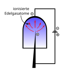
To examine a sample in a field ion microscope, a sharp metal tip is made and placed in a vacuum chamber filled with a noble gas (e.g. helium or neon ). Noble gases are used because you need a higher electric field to ionize them (closed shells). A higher electric field then also means higher resolution. The tip is cooled to a temperature between 20 and 100 K in order to reduce the unrest of the atoms due to Brownian molecular motion . A positive high voltage of 5 to 10 kV is applied between the tip and a detector, e.g. B. a combination of microchannel plate and phosphor screen applied. A negative voltage at the tip would cause undesirable field emission of electrons .
Gas atoms are ionized by the strong electric field near the tip (hence field ionization ), positively charged and repelled by the tip. The electric field strength must be sufficiently large to enable field ionization of the gas atoms, but it must be so low that detachment of atoms from the tip surface ( field evaporation or field desorption ) is avoided.
In contrast to other types of microscopes ( light microscope , electron microscope ), in which the magnification can be adjusted using optical elements (lenses), with the field ion microscope this essentially depends on the curvature of the tip surface and the voltage applied. The resolving power is not negatively affected by aberrations of optical elements. There is practically no wave-optical resolution limitation because of the low De Broglie wavelength of the ions. The ionized gas atoms are accelerated radially (perpendicular to the surface) from the tip surface onto the detector and convey a central projection of the tip surface with a magnification in the range of 10 5 … 10 6 . This makes it possible to map individual atoms of the tip surface without any problems and to remove lattice defects such as B. Observe dislocations .
See also
- Atomic probe
- In the case of the focused ion beam microscope , an ion beam generated as above is focused and used to examine an object.
- Surface chemistry
literature
- Erwin Wilhelm Müller : The field ion microscope , Z. Phys. 131 , 136 (1951)
