Bile duct
As a bile duct or bile duct to bile transport routes referred. There are intrahepatic (located in the liver , from the Greek hepar "liver") and extrahepatic (located outside the liver) bile ducts.
Intrahepatic bile ducts
The biliary tract begins with the biliferous canaliculi . The term biliary capillaries, which is also used, is misleading because, in comparison to blood capillaries, the biliferous canaliculi are not lined by an endothelium . Canaliculi biliferi are spaces between the liver cells without a wall of their own. They have a diameter of about 1 µm and are sealed by zonulae occludentes (liver-gallbladder barrier) . Cholangiocytes sitting in the wall of the bile tubules make up about a third of the bile, the rest comes from liver cells. The bile diffuses out of the liver lobule via the bile tubules in the direction of the respective Glisson's triangle .
Textbooks since 1959 have said that bile is already flowing in the canals. Bile salts would osmotically draw water from liver cells into bile ducts, which are only open towards the bile ducts. This is how a river is created. This has never been measured: biliary tubules are 100 times thinner than human hair. In 2020, intravital microscopy revealed that there is no measurable flow. It was also long assumed that the flow, which is stopped when the bile ducts constricts, builds up pressure that damages the liver. Drugs that lower the suspected flow should also relieve the damaging pressure. This was not aimed at the causes of fatty liver inflammation and has been refuted.
The bile canaliculi unite into a interlobular (between the lobules located) bile duct (ductus biliferus) , which together with a branch of the hepatic artery ( hepatic artery ) and portal vein (portal vein Vena) as so-called "Glisson's triad" (also called "liver triad" Trias hepatica ) runs in the periphery of a liver lobe. Bile ducts have a single-layer prismatic epithelium .
Extrahepatic bile ducts


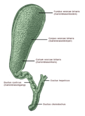
The extrahepatic biliary tract is used to drain and store the bile. For the right and left lobes of the liver, one hepatic duct emerges from the porta , the ductus hepaticus dexter and sinister . These combine at the hepatic fork to form the common hepatic duct (“common liver duct”, caliber in humans up to 7 mm; medical abbreviation often DHC). In some animals (for example predators , birds ) this union does not take place, so that no common liver duct is formed.
Anatomically very variable in height, but mostly in the middle third the ductus cysticus joins the ductus hepaticus.
The common duct after this union of the liver and gall bladder ducts is called the common bile duct . This opens on the papilla duodeni major with the ductus pancreaticus in the duodenum (duodenum) . The bile can thus be transported directly into the duodenum or initially for intermediate storage in the gallbladder.
Diseases
The diseases of the biliary tract include gallstone disease ( biliary concrements in cholelithiasis), inflammation (cholecystitis and cholangitis), functional disorders (dyskinesia, cholecystopathy) and tumors. Biliary atresia is a rare disease of newborns in which increasing inflammation of the extra- and intrahepatic bile ducts leads to a complete occlusion of the biliary tract. Bile duct cysts are rare malformations that are treated surgically. The hemobilia is bleeding into the bile ducts due to injury to a liver artery. The Cholangiocarcinoma is a malignant tumor of the bile duct.
Individual evidence
- ↑ N. Vartak, G. Guenther et al .: Intravital dynamic and correlative imaging reveals diffusion-dominated canalicular and flow-augmented ductular bile flux. , Hepatology June 19, 2020 https://doi.org/10.1002/hep.31422
- ^ Hans Adolf Kühn: Diseases of the biliary tract. In: Ludwig Heilmeyer (ed.): Textbook of internal medicine. Springer-Verlag, Berlin / Göttingen / Heidelberg 1955; 2nd edition ibid. 1961, pp. 875-885, here: p. 875 ( anatomical and physiological preliminary remarks ).
- ↑ Kühn, pp. 877 and 881-884.