Gallstone
| Classification according to ICD-10 | |
|---|---|
| K80 | Cholelithiasis |
| K80.0 | Gallbladder stone with acute cholecystitis |
| K80.1 | Gallbladder stone with other cholecystitis |
| K80.2 | Gallbladder stone without cholecystitis |
| K80.3 | Bile duct stone with cholangitis |
| K80.4 | Bile duct stone with cholecystitis Any condition listed under K80.5 with cholecystitis (with cholangitis) K80.5 |
| K80.5 | Bile duct stone without cholangitis or cholecystitis (clamped) gallstone: common bile duct common hepatic bile duct NOS Intrahepatic cholelithiasis hepatic colic (relapsing) |
| K80.8 | Other cholelithiasis |
| ICD-10 online (WHO version 2019) | |
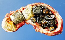
A gallstone , also called cholelite ( ancient Greek χολή chole , "bile", and λίθος líthos , "stone") or bile concrement , is a solid, crystallized precipitate of bile (bile). Gallstones are caused by an imbalance of soluble substances in the bile. If there is a gallstone in the gallbladder , it is called a gallbladder stone . If it is in the bile duct (common bile duct ), it is a bile duct stone. Related to gallstones, the gall semolina .
Generally, the presence of a gallstone is referred to as gallstone disease or cholelithiasis . More precisely, a gallbladder stone causes cholecystolithiasis (also cholecystolithiasis ; ancient Greek κύστις cystis , bladder) and a bile duct stone causes choledocholithiasis .
Gallstones are common and often cause no discomfort. However, if gallstones get stuck in the bile duct or in the gallbladder and obstruct the inflow or outflow of bile, severe colic and inflammation ( cholecystitis ) can occur.
Epidemiology
10 to 15 percent of the adult population are gallstone carriers, women are affected about twice as often as men. The disease is particularly common in western industrialized countries, and is even more common (60–70%) in the indigenous population of America. It occurs less frequently in East Asia, south of the Sahara and among African Americans. In Germany about half of the people have over 60 gallstones.
Genetic causes
In mid-2007, researchers at the Universities of Kiel and Bonn discovered a mutation in the ABCG8 gene as a cause of the formation of gallstones. (Three to four other mutations are suspected.) This gene contains the building instructions for steroline , which transports the blood fat cholesterol into the biliary tract. The mutation is said to increase this transport, which promotes the formation of gallstones. Research tries to inhibit the production of these proteins by introducing healthy genes or drugs and thus to find new approaches for prevention and therapy.
Emergence
The bile formed in the liver consists of the three main components bilirubin, cholesterol and the bile salts. With an imbalance of the soluble substances, accompanied by an inflammation or a flow obstruction in the biliary tract, for example due to a narrowing ( stenosis ) of the papilla (papillary stenosis ), stone formation can occur.
If there is an imbalance between bile acids and lecithin on the one hand and calcium carbonate or bilirubin on the other, calcium or bilirubin stones develop . If there is an oversupply of cholesterol and (more rarely) an undersupply of bile acids, cholesterol stones develop . 90% of the gallstones observed in Germany are cholesterol stones. However, it cannot be concluded from this that a particularly low-cholesterol diet could significantly reduce the risk of such stones forming. The administration of cholesterol-lowering drugs (statins) is therefore hardly suitable for significantly preventing gallstone formation.
In erythropoietic protoporphyria , stones often arise from protoporphyrin IX .
The development is promoted by the following factors:
- pregnancy
- family disposition
- Condition after small bowel surgery (bile acid loss syndrome)
- Diabetes mellitus (diabetes)
- Hypercholesterolemia (high cholesterol level)
- Hyperparathyroidism (disorder of the parathyroid glands)
- Crohn's disease (inflammatory bowel disease)
- Obesity ( obesity )
- High fat diet
- Chronic constipation ( constipation )
- Sedentary lifestyle
- Use of certain medications ( anti- ovulation drugs ( birth control pills ), clofibrate supplements)
- Hemolytic jaundice (jaundice)
- Rapid weight loss on a very low-fat diet
The Anglo-American also speaks of the “six Fs”: female, fat, fertile, forty, fair, family (female, overweight , fertile, forty years old, fair-skinned or blond , family accumulation).
Pathogenesis
The normal composition of bile is cholesterol , phospholipids and bile acids in a ratio of 1 to 5 to 14. Typical of lithogenic bile is the high proportion of cholesterol or the reduced proportion of bile acids, so that the bile is oversaturated with cholesterol. The following factors favor the formation of cholesterol gallstones:
- Gallbladder hypomotility , causing the bile to stay longer in the gallbladder
- incomplete emptying of the gallbladder
Symptoms
The gallstones only become symptomatic in about 25% of cases and therapy is only required in such cases. In this context, symptomatic means that colic occurs. These occur particularly after eating high-fat food or at night due to gallbladder contractions, when a stone z. B. clogged the output in the main bile duct. This leads to an increasing pain in the (right) upper abdomen, which can also radiate to the back or the right shoulder. In the case of a stone already in the bile duct, the bile builds up and the pressure develops more slowly up to the upper chest. This is also the case with an inflamed papilla.
Other symptoms:
- Tension pain in the (right) upper abdomen
- General symptoms of illness (bloating, flatulence, vomiting , sweating, loss of appetite )
- Jaundice ( jaundice ) due to increased leakage of bilirubin into the bloodstream
- brownish discoloration of the urine
- discolored, pale stool due to the lack of the blood breakdown product bilirubin
- increased liver values (e.g. Gamma GT)
diagnosis
In addition to the clinical examination, there are various apparatus-based examinations for diagnosis and confirmation of findings in the event of a suspected gallstone:

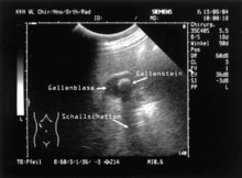
- Ultrasound examination ( sonography ): Gallstones and inflammation of the gallbladder can be shown well with ultrasound . However, small cholesterol stones that can clog the bile duct may not be found.
-
X-ray examinations :
- Blank image: Calcium-containing stones are shading in the X-ray image. Air in the gallbladder speaks for gas-forming pathogens.
-
Contrast medium examination :
- Oral cholecystography (replaced with ultrasound)
- Infusion cholecystangiography ("iv bile")
- ERCP (endoscopic retrograde cholangiopancreatography)
- MRCP (magnetic resonance imaging cholangiopancreatography)
- PTC (percutaneous transhepatic cholangiography)
- CT ( computed tomography )
- MRI ( magnetic resonance imaging )
- Laboratory diagnostics (detection of inflammation, differential diagnosis of jaundice)
treatment
Gallstones that do not cause symptoms do not require therapy. Colic requires abstinence from food , pain relievers, antispasmodics and, in the case of cholecystitis, antibiotics .
After the acute symptoms have subsided, there are several methods of removing the gallstones:
- Cholecystectomy : For stones in the gallbladder, an operation with removal of the gallbladder, either by laparoscopic or (less often) conventional surgery. The former is the therapy of choice, as it has a very low complication rate due to the minimally invasive procedure and definitely eliminates gallbladder stone disease. However, gallstones can remain in the bile duct or form anew and block it even after the gallbladder has been surgically removed. Then an ERCP is recommended. The removal of the gallbladder without a skin incision is offered as a new and not yet established procedure. In women this is done through the vagina, in men and women it is now also done through the stomach. This method is called NOTES (natural orifice transluminal endoscopic surgery).
- ERCP with stone extraction or expansion of the major duodenal papilla in the duodenum by means of papillotomy for stones trapped in the bile duct. It can be useful to do this before a cholecystectomy, as it is much less invasive.
- Litholysis: Drug dissolution of the stones with ursodeoxycholic acid (UDCA) or chenodeoxycholic acid. This method is only possible with pure cholesterol stones. Other prerequisites for lysis therapy are stone sizes of a maximum of 5 mm, whereby the gallbladder should not be more than half filled with stones, the absence of calcium deposits in the stones and the normal function of the gallbladder (after a "stimulating meal" the gallbladder should be at least 50 % empty, the bile ducts must be free). Contraindications are also acute or chronic inflammation of the gallbladder ( cholecystitis ) or the biliary tract ( cholangitis ), suspected gallbladder carcinoma , acute or chronic inflammation of the liver ( hepatitis ), pancreatitis ( pancreatitis ), liver cirrhosis , diarrheal diseases and pregnancy. Because of the frequent side effects of chenodeoxycholic acid, ursodeoxycholic acid, which is practically free of side effects, is preferred. Oral administration of ursodeoxycholic acid results in a stone-free condition of up to 70% within 1–2 years. The recurrence rate is up to 50% within 5 years. Intermittent, low-dose administration of ursodeoxycholic acid could be effective as prophylaxis. Therapy with ursodeoxycholic acid is the gentlest way to remove gallstones. The main advantages over an operation are the maintenance of a functional gallbladder, the avoidance of operational risks and their consequences and the maintenance of the ability to work during the treatment.
- ESWL (extracorporeal shock wave lithotripsy): Shattering the stones with shock waves, which then - usually with biliary colic - go off into the intestine. Here, too, there is a very high rate of recurrence, as new stones can form again in the gallbladder that is left in place.
Complications
- Colic
- Gall bladder hydrops with germ colonization and inflammation
- Acute cholecystitis
- Chronic cholecystitis
- Cholangitis
- Stone perforation - migration of a stone into the duodenum , stomach or colon (constriction Bauhin's valve ) with the risk of gallstone ileus
- Porcelain gallbladder (a precancerous condition )
- Biliary pancreatitis (inflammation of the pancreas) caused by a gallstone trapped in the papilla
New stone formation
Every 10th to 20th patient has at least one more colic after the gallbladder has been removed. This is mainly the result of remaining stones that had already got into the bile duct before or during the operation and were not removed with it. These can wander in the corridor, which slowly widens after the operation, and get caught in other places.
Even after the gallbladder has been completely removed, new gallstones can form in the bile duct. This affects about 1% of patients. This happens e.g. B. with a strong imbalance of the three main components of the bile. Bacteria that increasingly find their way into the bile duct, especially through today's use of modern endoscopic procedures, can promote gallstone formation. To treat such an acute stone, the Vater's papilla is cut open, for example with heating by electricity or it is smashed by means of ultrasound. If new stones appear again and again, the diet must be adjusted or medication intervened.
Connection with other diseases
In addition to the direct possible complications, a German study showed a 24 percent increased risk of cardiovascular disease in patients suffering from gallstones, which is probably due to the connection with the increased cholesterol. Removing the gallbladder did not reduce the risk, which is why previous gallstone patients should still be observed over the long term.
Web links
- Shaffer: EA Epidemiology of Gallstone Disease . PMID 17127183 (English)
- E-learning course on gallstones with images and videos (Charité Berlin)
- Ultrasound images of gallstones
- M. Frey, D. Criblez: Cholecystolithiasis . (PDF) In: Schweiz Med Forum , No. 32/33, August 15, 2001 (PDF; 229 kB).
- S3 - Guideline for the diagnosis and therapy of gallstones of the German Society for Digestive and Metabolic Diseases and the German Society for Visceral Surgery. In: AWMF online
Individual evidence
- ↑ Shaffer: Gallstone disease: Epidemiology of gallbladder stone disease . PMID 17127183
- ↑ Risk gene for gallstone discovered . Mirror online
- ↑ Genome-wide search identifies risk genes for gallstones . Science information service
- ↑ book u. a .: A genome-wide association scan identifies the hepatic cholesterol transporter ABCG8 as a susceptibility factor for human gallstone disease . In: Nat Genet . 2007 Aug, 39 (8), PMID 17632509 .
- ↑ Research team discovers gallstone gene . innovations-report.de
- ↑ MJ Khalili et al. a .: Erythropoietic protoporphyria and early onset of cholestasis. In: Turk J Pediatr. , 2012 Nov-Dec, 54 (6), pp. 645-650.
- ↑ a b Diagnostics and therapy of gallstones. ( Memento of the original from March 4, 2016 in the Internet Archive ; PDF; 1 MB) Info: The archive link was inserted automatically and has not yet been checked. Please check the original and archive link according to the instructions and then remove this notice. Guideline of the German Society for Digestive and Metabolic Diseases and the German Society for Visceral Surgery. AWMF Guideline Register No. 021/008. The validity of the guideline is currently being reviewed.
- ↑ Volker Hinz: Influence of ursodeoxycholic acid on the gastrointestinal quality of life index (GLQI) in patients with "asymptomatic" cholecystolithiasis . Dissertation . Ludwig Maximilians University, Munich 2005, DNB 975508407 .
- ↑ J. Wirth, R. d. Giuseppe et al. a .: Presence of gallstones and the risk of cardiovascular diseases: The EPIC-Germany cohort study. In: European Journal of Preventive Cardiology. March 2015, vol. 22, no. 3, pp. 326-334. doi: 10.1177 / 2047487313512218 .


