Gallbladder
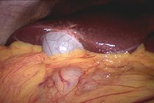
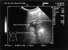
The gall bladder ( Vesica fellea or Vesica biliaris ; from Latin vesica "bladder" and fel or bilis "bile") is a hollow organ of vertebrates . In it the bile , which is produced by the liver for the digestion of fats in the intestines , is thickened and stored. Colloquially, the gallbladder itself is often referred to as "bile". Common diseases are of gallstones disabilities caused the supply and / or drainage of the gallbladder ( cholecystolithiasis and choledocholithiasis ) and triggered by gallstones inflammation of the gallbladder ( cholecystitis ). In humans, the gall bladder often has to be removed surgically ( cholecystectomy ). The most common examination method for assessing the gallbladder is sonography .
Occurrence
A gall bladder is formed in most vertebrates; it appears for the first time as a feature in the evolution of vertebrates . Within the vertebrate classes there are taxa that do not have a gallbladder. They found, for example in lampreys only in young animals as adults they will in the course of ontogenesis reduced. In addition, a number of cartilaginous fish do not have a gallbladder. Of the mammals , sloths , giraffes , tapirs , horses , rats, and deer do not have a gallbladder. Within the birds it is absent in most pigeons and parrots as well as the rhea and the African ostrich , in guinea fowl it is not always present. In the animal species without a gallbladder, the liver duct opens directly into the intestine (in mammals into the duodenum ).
Anatomical structure

9. Gallbladder, 10–11. Left and right lobes of the liver. 12. Spleen .
13. Esophagus . 14. Stomach . 15. Pancreas : 17. Ductus pancreaticus .
18. Small intestine : 19th duodenum , 20th jejunum
21-22: kidneys

The human gallbladder is usually 8 to 12 cm long and 4 to 5 cm wide. Their shape is often described as "pear-shaped". The gallbladder lies in the gallbladder fossa ( fossa vesicae biliaris ) on the underside of the liver between its quadrate lobe (square lobe) and dexter lobe (right lobe), but it can also be enclosed by liver tissue. In the caudal direction , the organ is related to the flexura coli dextra , the right bend of the large intestine ( colon ), which in the event of inflammation can lead to adhesions between the two organs or to connections between the respective cavities ( biliodigestive fistula ). Dorsally (backwards), the gallbladder is located medially in the immediate vicinity of the superior part of the duodenum ( duodenum ). In snakes , the gall bladder lies behind the liver and relatively far away from it.
The organ can be divided into a fundus vesicae biliaris (gallbladder floor ), corpus vesicae biliaris (gallbladder body) and collum vesicae biliaris (gallbladder neck ). The neck of the gallbladder, where the organ merges into the ductus cysticus (gallbladder duct ), has a spiral-shaped mucous membrane fold ( plica spiralis , also Heister valve ), which has a closing function, especially when increasing intra-abdominal pressure (e.g. when defecating ) perceives. The ductus cysticus unites with the ductus hepaticus communis to form the ductus choledochus , which runs in the ligamentum hepatoduodenale and opens into the duodenum.
The whole gall bladder is, with the exception of bodies which rest of the liver with peritoneum ( peritoneum coated), which from the right phrenic nerve innervated (phrenic nerve) sensitive. The nerve fibers originate from the spinal cord segments C3-C5. The fibers of the supraclavicular nerves , which innervate parts of the right shoulder, also originate from segments C3 and C4 on this side . If the peritoneum of the gallbladder is irritated by pathological processes, such as inflammation, this common origin can lead to the phenomenon of " transferred pain " in the shoulder. In addition, the gallbladder is vegetal fibers of the celiac plexus innervation.
The arteria cystica (bladder artery), usually a vessel from the right branch of the arteria hepatica propria ( liver artery ), supplies the gallbladder with blood . Laxative vessels are the venae cysticae, which open into the portal vein ( Vena portae ).
A variety known as the " Phrygian cap " is a bulging of the organ, the shape of which can be similar to that of the cap.
Feinbau

The approximately 0.4 cm thick wall of the hollow organ is histologically divided into three layers. From the inside (lumen) to the outside one differentiates between a tunica mucosa , consisting of epithelium and a lamina propria , a tunica muscularis and a tunica serosa .
The tunica mucosa ( mucous membrane ) consists of a layer of surface epithelium towards the lumen and an underlying connective tissue layer with blood vessels , the lamina propria . Due to the color of the bile , the mucous membrane is colored green. It is raised to wrinkles that are smoothed out as the filling increases. The accumulation of folds leads to so-called "mucosal bridges", which are characteristic of the histological preparation of a gallbladder. Occasionally occurring crypts are called Rokitansky-Aschoff crypts . The surface epithelium consists of so-called main cells, is single-layered and is characterized by a large number of microvilli . The cells are connected to one another by nexus , desmosomes and terminal ridges. The function of the main cells is to withdraw water to concentrate the bile and to produce mucus to protect the organ from bile components. In some mammals ( carnivores , ungulates ), the mucous membrane in the area of the gallbladder neck has mucous glands that synthesize mucins . With chronic inflammation, the number of these glands can be increased.
The middle of the three layers, the thin tunica muscularis , consists of smooth muscles in a concertina-like arrangement and isolated parts of connective tissue. The layer is necessary for the organ to empty.
The outer tunica serosa consists of the epithelium of the peritoneum and underlying connective tissue , with the exception of the area adjacent to the liver where a tunica adventitia is formed. In addition to nerve fibers , this layer also carries blood vessels.
Ontogenetic development
In ontogenesis , the development of the individual living being, the gall bladder emerges from a primitive intestinal tube, which is formed in the fourth week of development from the endoderm , the inner germ layer of the embryoblast . The cranial part of this tube (towards the skull) is called the foregut and is, among other things, the starting point for the development of the liver and gall bladder. The latter arises from the diverticulum cysticum , a protrusion of the foregut, which lies cranial to the anlage of the pancreas and caudal (towards the tail) of the liver anlage ( diverticulum hepaticum ). Both the gallbladder and the ductus cysticus (bile duct) develop from the diverticulum cysticum.
Both the absence ( aplasia ), the underdevelopment ( hypoplasia ) and the double structure of the organ are part of a multitude of rare malformations that are possible in humans. It is also possible to create direct passages from the liver to the gallbladder.
The gall bladder can be involved in rare syndromes , such as Mitchell-Riley syndrome .
physiology
The bile produced by the liver is used for digestion of fats in the intestine . The bile is released into the duodenum via the major duodenal papilla via the common bile duct . The sphincter muscles ( M. sphincter ampullae and M. sphincter ductus choledochi ) in the area of this opening can prevent the outflow of the bile by their contraction, so that it backs up into the gallbladder, which is connected via the ductus cysticus . This storage takes place mainly between meals ( interdigestive ) and affects about half of the bile secreted by the liver . The organ holds around 50 ml of bile, the concentration of which can, however, be greatly increased by actively withdrawing water. The bile can be enriched ("thickened") to up to ten percent of the original volume. In this context, the original “liver bile” is sometimes differentiated from the modified “bubble bile”. The latter is primarily characterized by an increased concentration of bile acids, lecithin , bile pigments and cholesterol . The thickening takes place by the displacement of sodium - and chloride ions using a Na + / H + - as well as a Cl - / HCO 3 - - antiport -Transportsystems in the apical (luminal) membrane of the main cell. This shift is electronically neutral, that is, no net charges are shifted. The water contained in the bile follows these resorbed ions due to their osmotic effectiveness . In the basolateral membrane of the cell there is a Na + / K + - ATPase , which keeps the intracellular sodium concentration constant. The absorbed water is transported away in the blood vessels of the lamina propria.
For relaxation ( relaxation ) of the sphincter it comes to the discharge of the contents of the gallbladder; This emptying is supported by the contraction of the smooth muscles of the gallbladder wall. The contraction takes place under the influence of cholecystokinin (CCK), the formation of which in the duodenum and upper jejunum ( empty intestine ) is stimulated, among other things, by fat in the food pulp, and the parasympathetic effect of the vagus nerve via the neurotransmitter acetylcholine .
Gallbladder diseases (cholecystopathies)
Gallstones are waste products from the bile. These calculi occur in around 12% of the German population, but only become symptomatic in around half of those affected. The causes can be, for example, an imbalance of the bile components bile acid and cholesterol . If bile acid is lost to a greater extent from the enterohepatic circulation due to insufficient absorption , as is the case with Crohn's disease , for example , or if it is insufficiently formed, the proportion of cholesterol increases relatively. This also applies to an increased level of cholesterol in the blood ( hypercholesterolemia ). Further substances subsequently accumulate on the crystallization core, which can lead to cholelithiasis (gallstone disease). Especially when very young people are severely affected, the cause can also be a build-up of the red blood pigment ( porphyria ), whose precursor products damage the bile duct cells.
Stone problems are often accompanied by pain in the abdomen, colic and jaundice . Treatment options include removal of the gallbladder ( cholecystectomy ) or removal, with or without crushing the stones, as part of an endoscopic retrograde cholangiopancreatography . A congestive gallbladder ( gallbladder hydrops ) occurs when the draining bile ducts are blocked by gallstones, stenoses or tumors while the production of mucins continues .
A common complication of gallstone disease is inflammation of the gallbladder ( cholecystitis ). It is a bacterial infection that is promoted in 90% of cases by a temporary obstruction of the gallbladder outlet. It can result in a build-up of pus in the hollow organ ( gallbladder empyema ). As a rule, if the gallbladder is inflamed, the organ must be surgically removed (usually as a laparoscopic cholecystectomy ). Recurring or chronic inflammation of the gallbladder can lead to a so-called "porcelain gallbladder", the wall of which calcifies due to the accumulation of calcium and thus hardens, or to a scarred "shrinking gallbladder ". The porcelain gallbladder in particular can prepare the ground for gallbladder carcinoma , a rather rare cancer with a poor prognosis.
The perforation of the gallbladder wall is called a gallbladder perforation or gallbladder rupture. This can be the result of cholecystitis as well as mechanical stress from a gallstone.
Around 5% of the population have gallbladder polyps . In the vast majority of cases, these are asymptomatic and benign; cancer is rarely hidden behind them.
Different as liver fluke called trematodes infect the biliary tree and gallbladder.
A biliodigestive anastomosis can be used to create an artificial connection between the gallbladder or the bile duct system and parts of the intestinal tract.
Investigation procedure
A healthy gallbladder is neither palpable nor tender. In the context of inflammation, it can be demarcated from the ventral edge of the liver if there is an accompanying enlargement, then there is usually a sensitivity to pressure during inhalation in the area of the costal arch ( Murphy's sign ). The organ can also be palpated with a bulging filling in connection with drainage disorders in combination with jaundice ( Courvoisier's sign ). This typically occurs when the excretory duct in the small intestine is blocked, for example by a pancreatic tumor .
A large number of methods are available for examining the gallbladder and biliary tract as well as possible pathological symptoms. Of these, sonography is the most popular because it is easy to perform and risk-free for the patient. Thus, sonography is the first procedure to assess the gallbladder, which may be followed by further examinations. The examination is usually carried out on an empty stomach, as the gallbladder is then filled and can best be assessed.
Other imaging methods for assessing the gallbladder, which are based on the X-ray display of the bile duct system and the gallbladder after administration of a contrast agent , are collectively referred to as cholangiography . Today it is customary to inject the contrast agent, which makes the organ visible, as part of an endoscopic retrograde cholangiopancreatography (ERCP) using an endoscope directly into the papilla duodeni major , the mouth of the bile duct system in the duodenum. This procedure enables not only the diagnosis of pathological changes such as gallstones or stenoses, but also the attempt of therapeutic intervention via the endoscope. If ERCP is not possible, percutaneous transhepatic cholangiography (PTC) is another option in which the contrast agent is introduced into the liver percutaneously , i.e. through the skin, by means of a puncture . Because of the advantages of ERCP, cholangiographies, in which a contrast agent excreted in the bile by the liver is administered as a tablet (oral cholecystography) or intravenously (intravenous cholecystography), have become uncommon today or are restricted to special indications.
The computed tomography is used in an unclear ultrasound findings, the spread diagnosis of tumors and to determine the lime content of gallbladder stones are used. An alternative to this is magnetic resonance tomography , which also allows the fluid-filled biliary tract, gall bladder and pancreatic duct to be reconstructed (magnetic resonance cholangiopancreatography, MRCP). In terms of its informative value, it is comparable to the ERCP and can be used if a therapeutic intervention is not planned in advance.
Due to the development of other diagnostic procedures, conventional X-rays have lost their place in the diagnosis of diseases of the gallbladder. The X-ray can be used to estimate the number and size of gallstones and identify a porcelain gallbladder.
Web links
literature
- Gerhard Aumüller, Jürgen Engele, Joachim Kirsch, Siegfried Mense; Markus Voll and Karl Wesker (illustrations): Anatomy , online learning program for the preparation course. 3. Edition. Thieme, Stuttgart 2014, ISBN 978-3-13-136043-4 .
- A. Benninghoff, D. Drenckhahn: Cell and tissue theory, development theory, skeletal and muscle systems, respiratory system, digestive system, urinary and genital system. 16th edition. Urban and Fischer, Munich 2003, ISBN 3-437-42340-1 (Anatomy, Volume 1).
- Renate Lüllmann-Rauch: pocket textbook histology . 4th edition. Thieme, Stuttgart 2012, ISBN 978-3-13-129244-5 .
- Thomas W. Sadler: Medical Embryology . Translated from the English by Ulrich Drews. 11th edition. Georg Thieme Verlag, Stuttgart 2008. ISBN 978-3-13-446611-9 .
- G. Skibbe: gallbladder and bile ducts. In: Surgery historically: beginning - development - differentiation. Edited by FX Sailer and FW Gierhake, Dustri-Verlag, Deisenhofen near Munich 1973, ISBN 3-87185-021-7 , pp. 72-88
Individual evidence
- ^ W. Westheide, R. Rieger: Vertebrate or skull animals. Spectrum, Heidelberg 2003, ISBN 3-8274-0900-4 (special zoology, part 2).
- ↑ a b Gerhard Aumüller et al .: Anatomie . 3. Edition. Thieme, Stuttgart 2014, ISBN 978-3-13-136043-4 , p. 667.
- ^ Gerhard Aumüller et al .: Anatomy . 3. Edition. Thieme, Stuttgart 2014, ISBN 978-3-13-136043-4 , p. 665.
- ↑ a b Gerhard Aumüller et al .: Anatomie . 3. Edition. Thieme, Stuttgart 2014, ISBN 978-3-13-136043-4 , p. 668.
- ↑ a b Renate Lüllmann-Rauch: Pocket textbook histology . 4th edition. Thieme, Stuttgart 2012, ISBN 978-3-13-129244-5 , p. 427 f.
- ↑ Thomas W. Sadler: Medical Embryology . Translated from the English by Ulrich Drews. 11th edition. Georg Thieme Verlag, Stuttgart 2008. ISBN 978-3-13-446611-9 , p. 287.
- ↑ a b Michael Gekle: nutrition, energy metabolism and digestion. In: Michael Gekle u. a .: Physiology. Thieme-Verlag, Stuttgart 2010, ISBN 978-3-13-144981-8 , pp. 461-463.
- ↑ Gerd Herold and colleagues: Internal Medicine 2018 . Self-published, Cologne 2018, ISBN 978-3-9814660-7-2 , p. 556 f.
- ↑ Gerd Herold and colleagues: Internal Medicine 2018 . Self-published, Cologne 2018, ISBN 978-3-9814660-7-2 , p. 570.
- ^ Maximilian Reiser, Fritz-Peter Kuhn, Jürgen Debus: Radiology. 3rd edition, Stuttgart 2011. p. 491.
- ^ Maximilian Reiser, Fritz-Peter Kuhn, Jürgen Debus: Radiology. 3rd edition, Stuttgart 2011. pp. 492 f.
- ^ A b Maximilian Reiser, Fritz-Peter Kuhn, Jürgen Debus: Radiology. 3rd edition, Stuttgart 2011. p. 490.
- ↑ Maximilian Reiser, Fritz-Peter Kuhn, Jürgen Debus: Radiology. 3rd edition, Stuttgart 2011. P. 493 f.


