Insect leg

Adult insects have 6 legs . All 3 thorax segments each carry a pair of legs. On Prothorax are the front legs, the Mesothorax the middle legs and the metathorax the hind legs.
construction

|

|
| Image 1: Scheme of the structure |
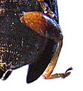
Image 2: Middle and hind legs of Procerus |
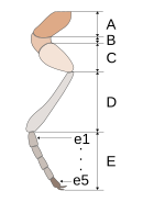
Each leg consists of 5 links (picture 1). Your name is:
- Α Hip ( Latin Coxa , plural: Coxae or Germanized also Coxen )
- B thigh ring (trochanter, from Greek trochós = disc, wheel; anteres = in front)
- C leg (Latin femur , plural: femora )
- D splint (Latin tibia , plural: Tibiae or Germanized also tibia )
-
E foot ( Tarsus , Greek. Tarsos = sole, plural: Tarsi or Germanized also Tarsen )
- The foot usually consists of 5 limbs (tarsomeres) (in the picture e1 ... e5) and an end piece.
- The first phalanx (e1) is also called the heel peg (metatarsus).
- The last foot link (e5) is also called the claw link (the term claw link is also used).
- On the end piece (pretarsus) there are claws and adhesive pads that are used to adhere to smooth surfaces.
In order to assign a limb to a pair of legs, the prefix of the thorax segment from which the pair of legs originates is placed in front of its name .
| Limb (general) | on the foreleg | on the middle leg | on the hind leg |
|---|---|---|---|
| Coxa | Procoxa | Mesocoxa | Metacoxa |
| Trochanter | Protrochanter | Mesotrochanteric | Metatrochanter |
| Femur | Profemur | Mesofemur | Metafemur |
| Tibia | Protibia | Mesotibia | Metatibia |
| tarsus | Protarsus | Mesotarsus | Metatarsus |
In the larval stages, the structure of the leg is sometimes very similar, but usually simpler, the legs can also be completely absent here. Insect larvae never have more than one tarsal link.
Notes on the leg links

|
|
| Image 3: Spotted shrimp | |

|

|
| Image 4a: Large birch sawfly |
Fig. 4b: Front rail of Ditomus |

Callidium violaceum
T splint (tibia)
K claws
1 tarsal limb 1
2 tarsal limb 2
For the tarsal links 3 to 5, see excerpt above:
tarsal link 3 - red bordered
tarsal link 4 - yellow
tarsal link 5 - green
The hip forms the connection to the part of the body called the chest. She is turned into a hip cavity. In beetles, this is called closed if there is a sclerite to the side of the abdomen (i.e. the hip is enclosed in a ring), otherwise open. The hip can be relatively large or small, round, cylindrical, conical (like the middle hip in Figure 2), straight, curved or flat (like the rear hip in Figure 2) and z. B. serve as a thigh cover. It can be barely visible in the body or, like the middle hip of the birch sawfly in Fig. 4a or the tentacles of the stick bug (Fig. 18), protrudes from the body like an “additional leg link ”.
The waist ring is usually a narrow ring that can be very small or missing entirely. In picture 2, the hip ring of the middle leg hugs the thigh and is much smaller than the hip ring of the hind leg, which protrudes backwards from the hind leg like an egg-shaped lump. In many insects it forms a joint that allows rotation. Hinge joints are formed between the hip and leg ring and between the leg and rail. The thigh ring and thigh are usually joined with a seam (suture), but without a joint.
The thigh and especially the splint often have teeth or thorns.
With the subordination of the long-feeler terrors, there is a hearing organ ( tympanic organ ) in the rail . In picture 3, the eardrum can be seen as a pale white oval in the upper section of the front splint of the spotted shrimp ( Leptophyes punctatissima ). On the thigh or the splint or on the body there are sculpted so-called shrill strips made of chitin , with which the insect can create sounds when it rubs them against each other.
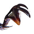
With some beetles you can find a so-called plaster notch (Fig. 4b). It consists of a small, round indentation on the inside of the rail that can be closed with a movable pin. She is surrounded by a series of short and stiff hair. These serve as a brush or comb for cleaning the sensor. When the beetle wants to clean its antennae, it brings its front leg closer to the head so that the antennae slides into the indentation of the rail. Then the thorn closes the opening, and when the animal removes the leg from the head again, the feeler brushes along the "brush". There is also a plaster notch on the bee , but this is on the first phalanx.
The foot members can have about the same size and shape as one another. B. in the hind tarsus of the beetle shown in Figure 7. However, they can also be designed very differently, this can be seen below with the bees . The ground beetles , which, as the name suggests, have good running ability, have 5 large tarsal links. The tarsi links can also be very small, a small one can “hide” in the appendages of a large one (Fig. 5) or there can be less than 5 tarsi links. In beetles, the number of tarsal limbs has taxonomic significance. The tarsi formula 5-4-4 means that the front legs each have 5 tarsi, the other legs only have 4 tarsi.
On the appendix of the last phalanx, the pretarsus, there are usually one or two claws, and very rarely both can be reduced. There may also be other attachments that are shown on the Faunistics website. The Prätarsus creates the connection to the underground and has to guarantee a secure adhesion. Their effectiveness becomes clear when observing a fly on the window pane or the ceiling. It has adhesive lobes between the claws, which are kept permanently moist by fine glandular hairs. Such detention devices are designed in various ways in different insect orders, they can be single or in pairs, sit on the claws or between them, occasionally also on the tarsal limbs themselves (e.g. in weevils and leaf beetles). In addition, the pretarsus also forms a sensory connection to the outside world of the insect. So on the underside of the Praetarsus there are also sensilles with the function of the sense of taste.
In the belly rings of the chest ( sternites ), depressions can be created that precisely match the thighs, splints or entire legs. So the insect can put these parts close to the body for protection, such as B. the ladybug .
Movement of the legs

|

|
| Fig. 6: Scheme | Figure 7: joint from the cricket |
The leg is part of the exoskeleton . The elements that give the leg strength and to which the muscles attach are not bones inside the leg, but rather through the hard shell of the leg, which essentially consists of chitin and proteins. The individual limbs of the leg are connected by softer joint membranes (Fig. 7). The muscles attach directly or indirectly via tendons from the inside to the exoskeleton.
The most important muscles are shown in Figure 6. The muscles that cause the limbs to flex towards each other through their tension are marked Y. Their opponents (antagonists), who cause the elongation when they are shortened, are marked with an X. Only bending and stretching are possible between the leg and the splint, the leg ring also allows turning. The corresponding muscle is marked with Z in Figure 2. The movement of the hip is different in the individual insect orders. There are no muscles in the tarsus. The muscles that move the claws are located in the thigh (picture 2, Y ') and in the splint and move the claws over a long tendon that runs through the entire tarsus.
The legs can have movable thorns, which are also connected to muscles.
The insects do not have blood vessels in their legs. The nutrients that are necessary for the functioning of the muscles are transported with the so-called hemolymph. With the hemolymph, which circulates freely in the body cavities, the waste products of muscle work are also removed. The muscle activity is controlled by the corresponding ganglia in the front, middle and rear chest.
Leg types

|
| Image 8: Head louse |
According to specialized types of locomotion, a distinction is made between the running leg, ankle leg and swimming leg. With the other types of muzzle, grave bone and collecting leg, the leg is each designed for a different function. It must be emphasized, however, that each leg performs several tasks and can be specialized beyond the “main purpose”.
The typification therefore only partially does justice to the abundance of leg shapes. An example of this are the legs of the lice (picture 8), which specialize in clinging to the hair. With them the strong claw of the Praetarsus forms a pair of pincers with an extension of the splint. There are also some species of beetle (Pogonostoma) that are so specialized in climbing trees that they can no longer move on the ground. The legs of dragonflies are very thorny and are used to catch prey, they are rarely or never used for running. Some species of ghosts , such as the Malay giant ghost, use their thorny hind legs to ward off enemies. For this purpose, the rails of the rear legs are quickly knocked against the thighs, which is a very effective defense due to the thorns in particular of the rails.
Running leg

|

|

|

|
||
| Photo 9: Middle legs of two tiger beetles |
Photo 10: Small cabbage white butterfly | Image 11: Schnake |
Of course, the leg's primary role is to enable the insect to move. The treadmill was developed for this, as can be found in beetles, especially in ground beetles (Carabidae) (Fig. 2). When walking, the thorny ends of the rails press into the ground, while the flattened feet rest on the ground. Some beetles, especially the tiger beetles (Cicindelinae), can walk very quickly. They have long and slim legs.
If beetles love to fly, their legs must be light. In Fig. 9 the legs of two different species of tiger beetle are shown, above a species that is less happy to fly, and below a species that is very happy to fly.
In insects that move almost exclusively by flies, such as butterflies (picture 10) or snakes (picture 11), the legs are, as expected, delicately built to save weight.
Ankle bone

|
|
| Figure 12: Short probe insect | |

|

|
| Photo 13: Flea beetle | Photo 14: Flea |
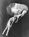
Jumping inevitably evolves from running fast, but has evolved into an independent way of moving. It is best known from grasshoppers (Fig. 12), but jumping was developed in many groups of insects. With regard to body size, this ability is particularly impressive in flea beetles (Halticinae, Fig. 13) and of course in fleas (Fig. 14).
All of these insects have thighs that are thickened because of the necessary jumping muscles. However, the reverse conclusion that thickened thighs indicate good jumping ability is wrong. Because the legs have other functions as part of the exoskeleton. For example, they can express the gender difference, as in the case of the soft beetles of the species Oedemera femorata , the males of which have thick thighs (Fig. 28), but the females do not have thighs.
Unusual shapes such as B. the sails on the back legs of the bug in Fig. 29 can serve as camouflage , deter or have other reasons. It is not always clear why natural selection favored the development of an unusual leg shape.
Swimming leg

|

|

|
|
| Fig. 15: Meso- and metatarsus of a piston water beetle |
Image 17a: hind leg of the jellyfish |

|

|
| Fig. 16: Fire beetle from below |
Image 17b: Hind legs tumbler beetle |
Another way of getting around is swimming. The rail, foot, or both take the form of an oar. Often rows of stiff hair form a widening of the “rudder blade”.
In picture 15 the legs of the large piston water beetle can be seen, above the middle and below the hind leg. In both of them, the paws form an oar that is broadened on the inside by a strip of hair. However, it is relatively little flattened and the last phalanx still has small claws. The hip still has limited mobility. A limited rotation around the axis is possible between the 1st and 2nd tarsal phalanx, which allows the broadside of the oar legs to be positioned in a streamlined manner during the swimming movement. The beetle lives in water, where it usually wanders around among the aquatic plants.
A better swimmer is the Gelbrand , whose hind legs are not only widened on the tarsi, but also on the tibia and on both sides by rows of hairs. The joint between the foot and the splint allows the tarsus to be rotated by 100 ° in an effective manner. The hip is immovably connected to the body, the joint to the trochanter is built in such a way that the thigh can only move parallel to the abdomen. The claws have disappeared on the hind legs (Fig. 16 and Fig. 17a). In order to hold on to aquatic plants, the Gelbrand uses the claws on the end link of the middle and front pair of legs.
The tumbler beetles of the genus Gyrinus move amazingly nimble on the surface of the water. The middle and rear legs are transformed into short and wide paddles (Fig. 17b) that move with a high flapping frequency (up to 50 times per second). The animals form groups whose members tirelessly whirl around like silver dots. Even among the bugs there are excellent swimmers with appropriately modified hind legs. Here the thigh, splint and tarsus are broadened by a row of hairs. Recently, the webbing hair system in back swimmers was scientifically investigated.
 
|

|
| Fig. 18A: Suckers on the foreleg of a male yellow beetle |
Fig. 18B: Praetarsus of the piston water beetle |

|
Photo 19: Water striders mating |
In aquatic insects that have developed swimming legs, there is also the development of smooth surfaces to improve flow behavior. However, the problem arises that the male has difficulties in mating to hold on to the female. Because of this, hair pads developed as suction cups on the protarsus of the males in some species. In Figure 18 A you can see the widened tarsi of the yellow-leaved male (left from above, right from below) with the suction cups on the left. In picture 18 B the protarsus of the male piston water beetle is shown, with which he can cling.
Another function of the legs in connection with water is to enable movement on the surface of the water. This has been achieved in excellent form with the water striders (Fig. 19).
Muzzle

|
 
|
 
|
| Photo 20: Catcher legs of the stick bug |
Picture 21: above praying mantis below water scorpion |
Image 22: Praying mantis mantis top from inside below from outside |
Predatory insects have front legs that specialize in grabbing prey.
The water scorpion ( Nepa cinerea ) spears its prey with the pointed, single-limbed tarsi (Fig. 21 below). Then he pricks her with the mouthparts and sucks her out. His hips are still relatively short.
The hips of the stick bug ( Ranatra linearis ), which also lives in water, are very long (Fig. 20). It doesn't have to be that close to the prey. With lightning-fast movements, the catch legs snap forward and then fold again, whereby the insect clamps the prey with the rough-grained surface of the rail and leg between them.
The catching horrors (picture 21 above) have even more impressive fangs . In picture 22 the inside and below the outside are shown above. You bridge an even greater distance with your long hips. The tarsus is only weakly developed. Rail and legs are equipped with sharp saw teeth on the side facing each other. In between there is no escape for the prey, it is eaten in peace by the praying mantis, regardless of its conspecifics.
Tombstone

|

|
| Image 23: Hermit's foreleg |

|
 
|
|
| Photo 24: Front leg grave runner from above (above) and below |
Image 25: Mole cricket , foreleg below |
What is impressive about some insects is their ability to dig. The shape of the front rail was transformed into a variety of shovels depending on the material being dug in.
The hermit ( Osmoderma eremita ) digs in loose gauze (Fig. 23).
The species of the ground beetle genus Scarites dig in moist sand (Fig. 24: front rail from above, front rail from below).
The mole cricket has a true multi-purpose digger (Fig. 25 above). Here the strongly widened leg is already designed in the shape of a shovel and is additionally provided with a curved hook with which hard earth can be broken up. The wide rail is also designed as a shovel and is also equipped with offset teeth with sharp edges. Any roots caught in it are cut off.
Collecting leg
 
|
|
| Image 27: Honeybee , above hind leg from inside below below hind leg from outside |
|

|

|
| Image 28: Mock longhorn beetle Oedemera flavipes |
Image 29: Anisocelis Passion Flower Bug |
The bees' collecting leg is well known (Fig. 27). The hind leg in particular is an organ used to collect and transport pollen and propolis. To do this, the splint and the heel link work together. The splint and the metatarsus are about the same size, the rest of the tarsi, on the other hand, are of normal width. The heel link looks like it belongs to the splint. The collection facility consists of four functional units.
The pollen basket is best known . Picture 27 below shows the hind leg of the bee from the outside. The small tarsal links can be seen on the left, followed by the broad and very hairy 1st tarsal link in the center left of the picture, which is rectangular to broadly oval. To the right of this is the equally wide rail that narrows towards the kinked leg. The flat and smooth rear rail is slightly arched towards the edges and forms the bottom of the pollen basket. The wall of the cup is formed by the long and stiff hair that surrounds the splint. Since the cup is strongly tilted to the side in a natural posture, the main load of the pollen lies on the part of the wall that borders the tarsus. As can be clearly seen in the picture, the hairs around the splint are longest there and run through each other so that they can stabilize into a braid when stressed. At the other edges of the splint, the hairs are parallel to each other and their tips are bent inwards when the basket is empty. When the basket is filled, they are gently pushed outwards and the bent tips hold the cargo together.
On the inside of the heel peg there are several parallel rows of parallel bristles, the so-called pollen brush, which can be seen as light-dark stripes at a small magnification in Figure 27 above. Furthermore, on the outside of the upper edge of the heel peg there is a blunt extension, the so-called pollen slider, at the top right of the first heel peg in Figure 27 and pointing downwards. Opposite it on the lower edge of the rail outside is finally the pollen comb, a wreath of short teeth. In picture 27 above they are dark against the light background, and light against the dark rail.
The pollen is partially moistened when the stamens are bitten open and after the bee rolls around in it, it sticks to the body hair. Since the bee has similar combs and brushes on the other legs as well, during the subsequent flight it can comb the pollen brushed from the body hair alternately out of the brushes and push it into the pollen basket with the pollen pusher.
Web links
Individual evidence
- ↑ Scheme and electron microscopic image of the Praetarsus ( Memento from August 29, 2008 in the Internet Archive )
- ↑ H. Freude, K. W. Harde, G. A. Lohse: Die Käfer Mitteleuropas, Vol. 9. Spectrum Academic Publishing House in Elsevier 1966, ISBN 3-8274-0683-8
- ↑ The beetles of the German Empire. Volume 1 (1908) Stuttgart, K. G. Lutz Verlag
- ↑ Bernhard Klausnitzer: Wonderful world of the beetles. Herder Verlag, Freiburg, ISBN 3-451-19630-1
- ↑ Construction of swimming legs and swimming movements in comparison with Dytiscus and Hydrophilus, English (PDF; 952 kB)
- ^ Bernhard Klausnitzer: Beetles in and on the water . ISBN 3-89432-478-3
- ↑ Summary of a work on the webbed hair system in back swimmers
- ↑ Recordings to the collecting leg
- ↑ Description of the bee's foraging behavior. DNB 974951056/34