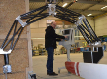
Medical C-arm
The medical C-arm is a medical product for imaging developed by Hugo Rost in 1954 . Characteristic of the medical C-arm of the C-shaped structure, the compound of is X-ray source and image receptor in which the position of two translational degrees of freedom (horizontal, vertical) and set two rotational degrees of freedom (orbital rotation, pivoting), and so the patient from almost every angle can be examined without moving it significantly.
history
The C-arm brought decisive advantages when it came to intraoperative imaging. Before him there was only conventional fluoroscopy and angiography for imaging e.g. B. during an operation. In both cases, however, the patient first had to be transported to the device, which meant further stress and waiting times for the patient and increasing costs for the hospital or practice. Only the mobile C-arm solved the problem of immobility and offered the option of moving the device to the patient if necessary.
Hugo Rost is the inventor of the medical C-arm, who developed the first C-arm together with Lothar Diethelm in 1954. After its introduction in 1955, the C-arm was initially used for X-ray diagnostics and enabled medicine to adopt new approaches to surgical planning.
Another milestone came with the introduction of compact high-frequency generators. With these, part of the generator and the X-ray tube of the C-arm could be accommodated in a common housing.
The emerging digitalization in medicine brought many advantages for the C-arm in the mid-1980s. The mere possibility of saving X-ray images on a computer and being able to call them up again at any time was an enormous relief for the medical staff. In addition, digitization was the starting signal for image processing programs, such as noise reduction to improve the recording quality and the subsequent digital subtraction angiography to improve the representation of vessels.
scope of application
Since its introduction in 1955, the C-arm has been continuously developed, which has made it into medical fields such as surgery , orthopedics , traumatology , vascular surgery and cardiology .
The C-arm is particularly suitable for intraoperative imaging. Here, every step of the operation can be monitored in real time, so that if necessary a revision can be carried out and a possible follow-up operation can be avoided, treatment results improved and recovery accelerated.
Another area of application can be found in vascular surgery. There, the C-arm is used to depict soft tissue and bone structures in detail. The C-arm is suitable for supporting biopsies and punctures , tumor embolization and ablation and drainage procedures .
The use of the C-arm is not limited to human medicine . It is also used in veterinary medicine and industry. In veterinary medicine it is used for intraoperative and minimally invasive interventions to obtain information during the operation. In industry, the C-arm is used in conjunction with laminographic processes for 3D imaging when analyzing very large test workpieces, e.g. B. Blades used in wind energy technology.
Individual evidence
- ↑ a b ARCADIS Orbic: Designed for Enhanced Surgical Precision. In: Center for Hip Health and Mobility. Vancouver Costal Health Research Institute, accessed March 16, 2020 .
- ↑ Christopher Nimsky, Barbara Carl: Historical, Current, and Future Intraoperative Imaging Modalities . In: Neurosurgery Clinics of North America . tape 28 , no. 4 , October 2017, ISSN 1042-3680 , p. 453-464 , doi : 10.1016 / j.nec.2017.05.001 .
- ↑ a b c Hugo-Rost & Co. GmbH history. Hugo Rost & Co. GmbH, accessed on March 17, 2020 .
- ^ Günther Stelzer: Surgical image intensifier . Ed .: MED - engineering. Issue 4, 2018, p. 60-61 .
- ↑ Tita, Ralf. Variable isocentric control for a standard C-arm with real-time 3D reconstruction . Dissertation Technical University of Munich, 2007.
- ↑ Laminography and Tomosynthesis. SHAKE GmbH, accessed on March 16, 2020 .

