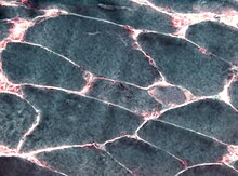
Muscle biopsy
The muscle biopsy is a procedure for the removal of muscle tissue that is used to diagnose muscle diseases ; mostly it is performed under local anesthesia .
Taking the muscle sample
The muscle sample can usually be taken on an outpatient basis. A muscle that has been affected by the disease but has not yet been completely destroyed by the disease is selected for the biopsy . When choosing a suitable muscle, magnetic resonance imaging or electromyography may be useful. However, the muscle should not have previously been damaged by electromyography electrodes or intramuscular injection . If inflammatory myopathy is suspected, the muscle biopsy should be performed before corticosteroid therapy is started, if possible.
Operational procedures
- In an open biopsy, a skin incision is made after local anesthesia and the muscle is exposed. A muscle tissue sample (approximately 0.5 cm³) is then removed. The wound is then closed with sutures after hemostasis. Complications during collection such as wound healing disorders or an infection are rare.
- The punch biopsy is less common, in which a small muscle tissue sample is removed through the skin after local anesthesia with a biopsy needle. Although this procedure is less invasive, only less muscle tissue can be obtained.
shipping
After the tissue has been removed, the tissue in a swab moistened with physiological saline solution is sent to an institute for neuropathology for histological processing . The transport takes place immediately by courier, as the further processing includes enzyme-histochemical staining , which can only be carried out on unfixed fresh material. To avoid freezing artifacts, the material is not frozen for shipping and is not shipped floating in liquid.
Neuropathological processing
In the neuropathological laboratory, after the sample has been oriented, sections of part of the tissue are first made on the cryostat in the transverse direction of the muscle fibers, with the remaining tissue being stored frozen in the event that Western blot tests are required. In addition, part of the sample is fixed and preserved in glutaraldehyde for any electron microscopic examinations that may become necessary . If there is residual material, it is fixed in formalin and embedded in paraffin.
Routine checkups

The neuropathological examination of the muscle sample routinely includes the following stains:
- Hematoxylin-eosin staining (including detection of inflammatory infiltrates, for example in myositis )
- Elastika van Gieson staining (EvG) (evidence of fibrosis of the endomysial connective tissue, e.g. in myopathies)
- Modified Gömöri trichrome staining (detection of inclusion bodies, for example in nemalin myopathy )
- PAS staining (evidence of increased storage of glycogen, for example in McArdle's disease )
- Oil-red color (evidence of increased lipid storage, for example in the case of a lack of carnitine palmitoyl transferase )
- Acid phosphatase reaction (evidence of increased macrophage activity, for example in inflammatory myopathies)
- NADH reaction (representation of the intermyofibrillary oxidative network and its disorders, e.g. in central core myopathy or multicore myopathy )
- ATPase reaction at different pH values (detection of the different fiber types and their disturbed distribution pattern, for example in the case of chronic neurogenic damage)
Further investigations
If muscular dystrophy is clinically suspected , immunohistochemical reactions, including antibodies against dystrophin and the sarcoglycans, follow. If there is no reaction, the results are validated in a specialized laboratory using Western Blot. The measurement of enzyme activities in metabolic myopathies is reserved for special laboratories, but can be carried out retrospectively on the preserved frozen material. Other special examinations, such as the in vitro contracture test to rule out malignant hyperthermia, can only be carried out in special laboratories on freshly removed tissue.
literature
- Victor Dubowitz, Caroline A. Sewry: Muscle Biopsy. A practical approach. Saunders, 2006. ISBN 1416025936
Web links
- Guide to the neuropathological diagnosis of muscle diseases (PDF; 106 kB) from the Reference Center for Neuromuscular Diseases at the DGNN

