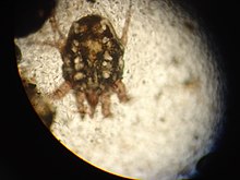
Ear mange
As ear mite ( otitis externa parasitaria ) refers to infection of the external ear and the external auditory canal with certain mites of the genus Psoroptes , Otodectes and Chorioptes . The corresponding representatives of these genera parasitize almost exclusively on the ear, only in the case of very severe infestation they can extend their infestation area to other parts of the head (head scabbard ) and body parts. On the other hand, other mange mites that do not specialize in the ear can also manifest on the ear, but the term “ear mange” is not common for this disease.
Pathogen
| Ear mite | infected species | Remarks |
|---|---|---|
| Cats | very often, rarely also transfer to humans ( zoonosis ) | |
| dogs | Rare | |
| marten | often in young animals kept in groups | |
| Red fox | ||
|
|
Rabbits | also with horses and ruminants ( goats , deer ) |
| Notoedres cati | Hedgehog | Pathogen that affects the cat's head |
| Chorioptes texanus | goat |
Clinical picture
An ear mange is usually associated with severe, bumpy skin changes on the inside of the auricle and the ear canal. The infestation with Otodectes cynotis causes thick, crumbly black-brown crusts, which result from the increased formation of ear wax and exudates as a reaction to the mites' saliva. In Psoroptes cuniculi , puff-like crusts dominate. Affected animals show strong restlessness and itching , which can lead to self-mutilation . A blood ear (othematoma) can develop in dogs .
As a complication, the ear infection can break through the eardrum and spread to the middle and inner ear , the meninges or the brain . A bacterial secondary infection of damaged skin of the ear canal is frequent.
The diagnosis can be made with an otoscope , with which the mites can be seen as dark, moving points in the ear canal. The diagnosis can be confirmed with a microscopic examination of a smear from the ear. What kind of mite it is can also be seen under the microscope.
treatment
Treatment is done by thoroughly cleaning the ears and removing the crusts. The mite control is carried out using acaricidal agents such as ivermectin (not in dogs with MDR1 defects ), doramectin or selamectin . The active ingredients are often administered via so-called spot-on preparations, which are dripped into the neck of the animal. Ivermectin can be administered both locally and systemically; the other two avermectins are administered systemically. Oral treatment with Fluralaner is also possible. Fipronil is also effective when applied locally , but the active substance should not be used in rabbits. Antibiotic ear drops are administered to counteract the secondary bacterial infection. Other dogs and cats living in the household should also be treated in order to prevent mutual infection. With consistent treatment, the prognosis for ear mites in dogs is favorable.
Individual evidence
- ↑ Van de Heyning, J .; D. Thienpont: Otitis externa in man caused by the mite Otodectes cynotis. Laryngoscope 1977, 1938-1941.
- ↑ Wieland Beck: Ear mange by Otodectes cynotis (Acari: Psoroptidae) in ferrets - pathogen biology, pathogenesis, clinical features, diagnosis and therapy. In: Kleintierpraxis 46 (2001), pp. 31–34.
- ↑ Josef Boch & Rudolf Supperer: Veterinärmedizinische Parasitologie Georg Thieme Verlag, 2006, pp. 542-544.
- ↑ G. Sheinberg et al .: Use of oral fluralaner for the treatment of Psoroptes cuniculi in 15 naturally infested rabbits. In: Vet. Dermatol. Volume 28, 2017, pp. 393-395.
- ↑ Curtis, CF: Current trends in the treatment od Sarcoptes, Cheyletiella and Otodectes mite infestations in dog and cats. Vet. Dermacol. 15/2004, pp. 108-114.
- ↑ Wieland Beck: Ear mange due to Psoroptes cuniculi (Acari: Psoroptidae) in domestic rabbits - pathogen biology, pathogenesis, clinical features, diagnosis and therapy. In: Kleintierpracxis 45 (2000), pp. 301-308.
- ^ Ernst G. Grünbaum & Ernst Schimke: Klinik der Hundekrankheiten Georg Thieme Verlag, 2005, p. 252.


