Photoluminescence spectroscopy
In the photoluminescence - spectroscopy (PL) spectroscopy is brought to the material under investigation by light absorption in electronically excited energy states which then under emission of light (spontaneous photon emission in the form of fluorescence or phosphorescence ) again reaches energetically lower lying energy states. The emitted light is detected and provides information about the electronic structure of the material.
Photoluminescence spectroscopy is a very sensitive method to investigate both intrinsic and defect-related electronic transitions in semiconductors and insulators . Optically active defects can be detected in concentrations of up to . While the method was primarily used in basic research at first , it is increasingly being used industrially for non-destructive, spatially resolved material characterization due to the steadily increasing demand for highly pure or specifically doped materials .
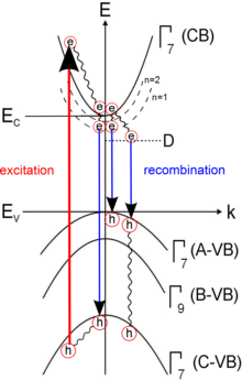
In the photoluminescence of semiconductors are by excitation with photons with electrons from the valence band into the conduction band lifted. These excited charge carriers thermalize in a non-radiative manner to the band edges of the conduction and valence bands by releasing the energy to the semiconductor crystal in the form of phonons . Depending on the crystal, these free charge carriers recombine as electron-hole pairs , the so-called excitons, or fall into lower-lying states and recombine from there. With PL spectroscopy, free excitons, excitons bound to defects, donor-acceptor pairs and other states that result from higher levels of the exciton (n = 1,2, ...) or from combinations of holes can be identified form different valence bands (A, B or C). Donors, acceptors and crystal defects such as dislocations come into consideration as impurities . These states then recombine with characteristic lifetimes, which enable detection (e.g. via time-resolved photoluminescence spectroscopy (TRPL, time-resolved PL)). The energy released can be emitted in various forms, for example as phonons via the crystal lattice , as photons or as Auger electrons . In photoluminescence, the photons emitted by radiating recombination mechanisms are detected. If several excited states exist, generally only transitions from the lowest state can be observed due to the very rapid thermalization. The measured radiation allows important insights into the properties of the examined substance, for example into the defect inventory of the crystal and its band gap .
Most photoluminescence measurements are carried out at low temperatures (sample cooled by liquid nitrogen , 77 K, or helium , 4 K,) in order to prevent thermal ionization of the optical centers and to avoid the broadening of the photoluminescence lines by lattice vibrations (phonons) . At low temperatures, most of the excitons are in a bound state and thus enable statements to be made about the energetic position of the impurities and the concentration of impurities.
Experimental setup
The luminescence of the material to be examined is achieved by light excitation, for example by a laser of sufficient energy. In general, a filter is placed in front of the laser that eliminates non-lasing lines of the laser plasma that lie in the relevant spectral range. The laser beam is focused on the sample in the cryostat through a lens . The emitted light (and partly also scattered light from the laser) is in turn focused by 2 lenses onto the entrance slit of the monochromator . Ideally, a filter is installed in front of the gap to filter the scattered laser light. In the monochromator, the radiation is spectrally split by variable grids (can be selected depending on the desired wavelength range ) and the dispersed light is directed onto the detector. There the optical signal is converted into a current or voltage signal, depending on the type of detector. In the case of amplification and phase-sensitive detection of the signal using a lock-in amplifier , the radiation (ideally right in front of the radiation source) must be modulated by a chopper .


