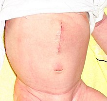
Pyloric stenosis
| Classification according to ICD-10 | |
|---|---|
| K31.1 | Hypertrophic pyloric stenosis in adults |
| K31.3 | Pylorospasm, not elsewhere classified |
| Q40.0 | Congenital hypertrophic pyloric stenosis |
| ICD-10 online (WHO version 2019) | |
The pyloric stenosis or Pförtnerverengerung describes a narrowing in the area of pyloric . This can be congenital or acquired. It leads to a disturbed transmission of the stomach contents into the duodenum and thus to insatiable vomiting . Treatment usually consists of surgical correction of the bottleneck.
Congenital pyloric stenosis
When stomach cramp porter (Pylorusmyohypertrophie; Engl .: Pylorospasm ) opens the pylorus , the muscle that closes the stomach to the duodenum not permanent and can not be the stomach contents to pass more. The persistent cramping causes the muscle to thicken over time.
Occurrence and causes
The disease starts at birth and occurs in families (possibly hereditary). The causes have not yet been clarified. The disease is found mainly in Western and Northern Europeans with a frequency of 1: 300, rarely in Asians and almost never in Africans. The peak of the disease is three weeks after birth. The disease occurs particularly in the first-born boys (ratio boys: girls: 4-5: 1).
Symptoms
The infant vomits (not biliously) about half an hour after the meal, partially or completely, like a surge. The stomach irritation may have threads of blood in the vomit. Then he looks for food again. Immediately after a meal, increased stomach movements ( peristalsis ) can be observed on the abdominal surface in the upper abdomen. The enlarged pylorus is partially palpable. The affected children are malnourished, underweight, constantly hungry and correspondingly dissatisfied due to the obstructed food passage. They get rid of low-mass starving stools at high frequency.
Risks
The lack of food intake and repeated vomiting can lead to pronounced metabolic disorders (e.g. "coma pyloricum") and dehydration. It is therefore often necessary to supply fluids by infusion.
diagnosis
The diagnosis is made on the basis of symptoms and ultrasound , which makes the hypertrophic pylorus muscle visible. As a result of vomiting, there is a chloride deficiency due to the loss of gastric juice , which leads to a disturbance of the acid-base balance in the sense of metabolic alkalosis . Due to alkalosis, more potassium is absorbed from the blood into cells, causing hypokalemia .
X-ray diagnostics can be performed in clinically unclear cases . A large gastric blister is evident on the abdominal survey. The diagnosis to differentiate similar diseases can be confirmed by gastrointestinal passage of contrast medium .
therapy
Conservative
The dehydration and alkalosis, which are common in chronic vomiting, are corrected until the final operative therapy . Furthermore, antispasmodics and small meals.
Operational
The pyloric stenosis is almost exclusively treated surgically. The gatekeeper is split lengthways down to the mucosa , which remains intact (pyloromyotomy according to Weber- Ramstedt ). Just a few hours after the operation, the baby can be used to normal nutrition again.
Differential diagnosis
Other changes with obstruction of gastric emptying are to be distinguished, e.g. B. duodenal obstruction , in the context of malrotation of the intestine or, less commonly, a pyloric atresia . A disease with similar symptoms is adrenogenital syndrome (AGS). However, the vomiting is mostly "flaccid". In contrast to pyloric stenosis, however, the potassium level in the blood in AGS is normal or increased.
Acquired pyloric stenosis
causes
Acquired pyloric stenosis can result from inflammation, gastric or duodenal ulcers or tumors of the stomach and adjacent organs. As a rule, however, it arises idiopathically, ie without an identifiable cause. There is a significantly higher incidence in boys, so a genetic component is likely.
Symptoms
Porridge stasis, fetus , vomiting, desiccosis , hypochloraemic alkalosis , marked gastric peristalsis, weakness, marasmus or cachexia
diagnosis
The diagnosis is usually made on the basis of the very typical symptom constellation and a subsequent ultrasound examination. This involves measuring the wall thickness and the length of the pylorus and assessing the peristalsis in the stomach, which, in the presence of pyloric hypertrophy, shows a retrograde movement of the chyme in the stomach. In addition, a determination of the electrolytes in the blood is carried out, whereby the chloride level and the excess of base are decisive. If a secondary cause comes into consideration, gastroduodenoscopy ( gastroscopy ) or another imaging method should often be selected, preferably magnetic resonance imaging .
therapy
Depending on the underlying disease, different surgical procedures are available ( pyloroplasty , resections, gastrectomy, cephalic duodenopancreatectomy ).
See also
- Roviralta syndrome , a combination of pyloric stenosis and hiatal hernia .
Individual evidence
- ^ Pschyrembel: Clinical Dictionary Walter de Gruyter, 265th edition (2014), ISBN 3-11-018534-2 .
- ^ V. Hofmann, KH Deeg, PF Hoyer: Ultrasound diagnostics in paediatrics and pediatric surgery. Textbook and atlas. Thieme 2005, ISBN 3-13-100953-5 .
- ↑ M. Bettex, N. Genton, M. Stockmann (Ed.): Pediatric surgery. Diagnostics, indication, therapy, prognosis. 2nd edition, Thieme (1982), ISBN 3-13-338102-4 .
- ↑ W. Schuster, D. Färber (Ed.): Children's radiology. Imaging diagnostics. Springer 1996, ISBN 3-540-60224-0 .
literature
- S1 guidelines for hypertrophic pyloric stenosis of the German Society for Pediatric Surgery (DGKCH). In: AWMF online (as of 2013)
- FC Sitzmann: Pediatrics. Diagnostics - therapy - prophylaxis. 6th edition, Hippocrates 1988, ISBN 3-7773-0827-7
- Hans Adolf Kühn: Diseases of the stomach and duodenum. In: Ludwig Heilmeyer (ed.): Textbook of internal medicine. Springer-Verlag, Berlin / Göttingen / Heidelberg 1955; 2nd edition, ibid. 1961, pp. 767-804, here: pp. 796-798.

