Temporal lobe


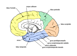
The temporal lobe ( Latin tempus 'temple' ) or temple lobe (Latin lobus temporalis ) is one of the four lobes of the cerebrum and makes up its laterobasal (lateral and lower) part. He is upward and forward from the lateral sulcus ( Sylvian fissure ) against the parietal lobe ( parietal lobe , parietal lobe ) and the frontal lobe ( frontal lobe , frontal lobe defined), to the rear it borders on the occipital lobes ( occipital lobe , occipital lobe ). The temporal lobe contains the primary auditory cortex , the Wernicke language center and important structures for memory .
Function and location
Primary auditory cortex
In the depth of the lateral sulcus lie the transverse temporal gyri (Heschl's transverse turns, named after the Austrian anatomist Richard Heschl (1824–1881)), which form the primary auditory cortex ( Brodmann area 41). After crossing the ear, the nerve fibers run from the ear to the opposite side via the lemniscus lateralis to the colliculi inferiores and from there via the pedunculus colliculi inferioris to the corpus geniculatum mediale . They then reach the primary auditory cortex through the internal capsule . Analogous to the sensory and motor cortex, the auditory cortex shows a tonotopic structure according to frequencies. The auditory pathway has several commissure systems that allow fibers to be changed. In this way, some fiber bundles also reach the auditory cortex on the same side, which is important for directional hearing.
Wernicke Language Center
In the superior temporal gyrus there is a sensory language center (the so-called Wernicke center , Brodmann area A22), which is important for language understanding. A whole series of other areas are involved in the processing and understanding of language. The Wernicke area occurs only in the dominant cerebral hemisphere (that is, in the hemisphere in which language is processed both motor and sensory), which is usually located on the left in right-handers, but is optionally on the left or right in left-handers can.
Memory structures
The medial part of the temporal lobe contains the hippocampus , the entorhinal , perirhinal and parahippocampal cortex , which represent the most important "coordination center" of declarative (explicit) memory .
The lower temporal area (IT) itself is part of the visual working memory ( working memory ). What is currently being perceived is briefly saved here (seconds to minutes). The comparison with the next perceptual content is also made here. The hippocampus is only needed if certain information is to be remembered in the medium to long term.
Neocortical associative areas
Many areas of the temporal lobe are also responsible for the recognition of complex non-spatial auditory and visual stimuli, such as B. the recognition of body parts - especially faces - and other meaningful objects (food, prey).
With electrical stimulation of the IT (experiments by Wilder Penfield 1959 in epileptic patients), event hallucinations ( experiential response ) occur, which the patients describe as coherent memories of past experiences. These memory reactions could only be generated when the temporal lobe was stimulated; they were rather rare (8% of the stimulus attempts) and their origin and meaning are not yet clear.
Blood supply to the temporal lobe
The basal and posterior medial part of the temporal lobe is supplied by the arteriae cerebri posteriores , which arise from the arteria basilaris . The lateral and anterior medial part receives its blood supply via branches of the arteriae cerebri mediae from the arteria carotis interna . The blood drainage takes place via the descending superficial veins of the brain ( Venae superficiales descendentes cerebri ) and via the middle superficial cerebral vein ( Vena media superficialis cerebri ). The descending veins drain into the transverse sinus . The drains of the central vein go both in the cavernous sinus , and in the transverse sinus . The transverse sinus eventually directs blood to the internal jugular vein that leads out of the skull.
pathology
Corresponding to the diverse functional areas of the temporal lobe, multiple disorders and impairments occur in traumatic or iatrogenic lesions.
The first and best-studied case was the assembly line worker Henry Gustav Molaison , who had the medial areas of both temporal lobes removed due to an untreatable epilepsy . After the operation, the patient suffered from severe anterograde amnesia and was no longer able to transfer what he had learned into long-term memory. He showed a normal command of the language and his vocabulary and intelligence quotient remained in the slightly above-average range. Due to the memory loss, he was no longer capable of explicit learning. However, he was still very capable of implicit learning even if he wasn't aware of it.
Lesions of the associative temporal cortex can lead to multiple visual and auditory deficits ( agnosia ). The recognition or naming of objects can be disturbed (object agnosia) or the recognition of faces ( prosopagnosia ). Auditory agnosias concern the inability to recognize tones, melodies, rhythms or tempos of music ( amusia ). Furthermore, word and language comprehension disorders ( aphasia ) can occur if the upper left and middle area of the temporal lobe is affected ( Wernicke area ).
See also
literature
- Eric R. Kandel et al. a. (Ed.): Neurosciences. An introduction . Spectrum Akademischer-Verlag, Heidelberg 1996, ISBN 3-86025-391-3 .
- Gerhard Roth : The brain and its reality . Suhrkamp, Frankfurt / M. 2002, ISBN 3-518-28875-X .
- Johannes Sobotta (ed.): Atlas of the human anatomy . Elsevier, Urban & Schwarzenberg, Munich 2004, ISBN 3-437-43590-6 .
Web links
- Christian Siedentopf: Page about fMRI
Individual evidence
- ↑ Federative Committee on Anatomical Terminology (Ed.): Terminologia Anatomica . Thieme, Stuttgart 1998 (English).

