Family tree analysis

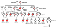
The analysis of family trees , like twin research and analyzes using the population statistical method, is one of the oldest methods of genetic research in humans. Since the discovery of chromosomes , the analysis of karyograms has been added. Knowledge of Mendel's rules is a prerequisite for understanding . The method of family tree analysis was developed especially on the basis of the inheritance of visible hereditary diseases . The observable different inheritance patterns , however, can be found in all hereditary characteristics inherited through chromosomes, i.e. also in most of the things that make up the healthy overall organism.
Procedure for family tree analysis for genetic traits that are encoded on the DNA in the cell nuclei :
- Is the characteristic under consideration inherited dominantly (pronounced in every generation) or recessively ?
- Is the characteristic under consideration inherited as an autosomal or gonosomal (one gender particularly affected)?
- Precise details of each safe and possible genotype as well as the reason for it (for example by means of cross-breeding schemes ).
It is also important to justify why the respective alternative was excluded.
Signs of an autosomal dominant inheritance

In the case of an autosomal dominant inheritance , a heterozygous individual can also be recognized by the phenotype . Here a heterozygous carrier is sufficient to produce an average of 50% offspring affected by the characteristic with a partner who does not have the characteristic, i.e. who is definitely homozygous and healthy, who are then heterozygous again and show the characteristic phenotypically. It can also happen that there are no affected offspring because the alleles in oogenesis and spermatogenesis reach the gametes more or less randomly ( meiotic drive ), but then the characteristic does not reappear in the following generations with healthy partners because in this case only the healthy recessive allele was passed on. In the case of those individuals who do not show the dominant trait within a dominant-recessive inheritance , one can assume that they are homozygous and do not inherit it. The proportion of affected offspring can also be higher than 50%, depending on how the two alleles are distributed during gamete formation.
The inheritance scheme for the connection of two heterozygous carriers of a dominant trait corresponds to Mendel's rule of splitting . If two heterozygous partners produce offspring together, an average of 75% of them become carriers of traits, with one third being homozygous and two thirds being heterozygous. You can't tell the difference between these two by the phenotype. You can see an increased occurrence of the trait, and only the remaining, on average, 25% do not show the trait because they each received the recessive allele from both parents, i.e. they are homozygous. The distribution of a trait between the two sexes is roughly the same for autosomal inheritance, provided that a sufficient total number of individuals is entered in the family tree so that a statistical mean can be formed on this basis.
Signs of an autosomal recessive inheritance

If two phenotypically healthy parents have children affected by an autosomal inherited disease, the conclusion can be drawn that the disease-causing gene is inherited recessively . In this case , both parents are conductors , i.e. carriers of the hereditary disease without being affected themselves. Your genotype is heterozygous . Inheritance takes place according to the split rule .
Only those children who have received the recessive allele from both parents, i.e. who are homozygous , become ill. Those children who received the recessive allele from only one parent are healthy themselves, but are conductors and can therefore pass on the genetic make-up. The children who received only the healthy allele from each parent are genetically healthy and therefore cannot inherit the disease.
In a pedigree analysis, an autosomal recessive inheritance can be inferred if the proportion of the sexes is the same among those affected (i.e. gender- independent inheritance) and if it also happens that one or more generations are skipped at individual points in individual lineages "will only appear again in the following generation. Sometimes there can be no more sick people at all in the following generations, if only connections with homozygous healthy partners have been established. The sequence in which the phenotypes appear allows conclusions to be drawn about the genotypes of individual individuals, although these are not always clear.
Characteristics of gonosomal inheritance
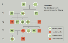
In most cases of gonosomal inheritance , the affected alleles are on the X chromosome ( X-linked inheritance ). However, there are also a few (few) exceptions where the allele in question is on the Y chromosome ( Y-chromosomal inheritance ). However, this form of gonosomal inheritance is only found very rarely because the Y chromosome is largely empty and is therefore irrelevant for the inheritance of traits . Typical examples of X-linked modes of inheritance are the inheritance of hemophilia and the red-green color blindness , in which the affected allele is on the X chromosome, with women, so a gene defect on one have its two alleles, female carriers are , i.e. not showing the trait in the phenotype .

- In a gonosomal - recessive inheritance more men than women are affected by the hereditary disease significantly, since men have only one X chromosome have, the Y chromosome can not oppose, because it is not really homologous chromosome. Therefore, when a male child is conceived, an allele on his X chromosome already affects the phenotype (hereditary scheme see carrier ). In girls, the recessive allele would have to be present on both chromosomes in order to have an effect on the phenotype. Only if a man who is affected himself fathered a daughter with a female carrier, there is a 50% probability that she will also be affected, depending on whether she receives the dominant healthy or the recessive disease-causing allele from the mother. In the case of blood clotting disorders, there are also rare autosomal inherited genetic defects that also lead to haemophilia . With them, the proportion of the sexes is the same among those affected. The hemophilia has life-threatening effects in girls from the onset of menstruation .
- In a gonosomal - dominant inheritance, in which the disease-causing allele is on the X chromosome, the daughter of a man concerned is also affected because the disclosure of his X chromosome in spermatogenesis causes it is a girl. Fathers only pass the allele on to their daughters, as male offspring only inherit the information on the father's Y chromosome from the father. An inheritance of the corresponding trait from father to son is therefore not biologically possible. This means that if a son is still affected, the disease-causing allele must be on the X chromosome from the mother.
Maternal inheritance
Genetic traits inherited through mitochondrial DNA usually follow a maternal inheritance, i.e. are only inherited from the mother, because the zygote only contains the mitochondria of the egg cell. The mitochondrion of the sperm usually does not get into the zygote. Women affected by mutations in the mtDNA pass the trait on to their children of both sexes. Affected men do not pass it on to any of their children.
Efficiency of the method
Using these criteria, family trees in humans and also pedigrees in breeding animals that go over several generations and whose entries are correct and complete can be analyzed relatively precisely. From the way in which a characteristic occurs in the generation sequence, one can infer the genotype of individual individuals. This method is only suitable for inheritance in monogenic characteristics. It should also be taken into account that conductors with rare hereditary factors can also remain undetected, as long as they do not accidentally produce offspring with other conductors. For this reason, when the method of family tree analysis was developed, it was precisely those family trees in which hereditary diseases were visible that were of interest. Modern molecular and human genetics now have even more efficient methods, although family tree analyzes are still carried out in advance of other investigations, as this provides important clues as to which genetic loci might need to be examined.
See also
Web links
Individual evidence
- ^ School of Biology: Family Tree Analysis
- ↑ Karl Skorecki, Doron Behar: Mitochondrial DNA and hereditary features and diseases
- ↑ Dr. Carmen L. Battaglia: Pedigree Analysis
- ^ Neil A. Campbell , Jane B. Reece : Biology. Spektrum-Verlag 2003, ISBN 3-8274-1352-4 , pages 308-311.
- ^ Ulrich Weber: Biologie Gesamtband Oberstufe, Cornelsen-Verlag 2001, ISBN 3-464-04279-0 , pages 178-182.
