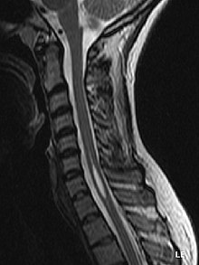
Syringomyelia
| Classification according to ICD-10 | |
|---|---|
| G95.0 | Syringomyelia and Syringobulbie |
| ICD-10 online (WHO version 2019) | |
The syringomyelia (from ancient Greek σύριγξ syrinx , German , pipe, flute ' and νωτιαῖος μύελος nōtiaios mýelos , German , spinal cord' ) is a formation of a cavity in the gray matter of the spinal cord . Synonym is also hydromyelia (of ὕδωρ hydor , German , Water ' ) or the combination of both concepts of Hydrosyringomyelie spoken.
The cervical and thoracic marrow is often affected; if it is located in the medulla oblongata , it is called syringobulbie .
Epidemiology
It's a rare condition. The prevalence is 8.4 per 100,000 inhabitants, although other studies show different prevalence rates (e.g. 1.94 per 100,000 inhabitants in Japan). In Germany, the incidence of syringomyelia varies from region to region: spinal cord disease is more common in southwest Germany and Austria.
Men are more often affected than women. The disease typically occurs in late puberty or young adulthood (between the ages of 20 and 40).
With the increased and improved imaging by magnetic resonance imaging, more and more cases of asymptomatic syringomyelia are being discovered. An epidemiological study from Japan on the prevalence of syringomyelia concluded that 22.7% of all syringomyelia do not cause any symptoms.
causes
There are different theories about the development of syringomyelia, whereby the theories have to be differentiated according to the form of the disease. A non-exhaustive list of theories of origin is given below:
- Hydrodynamic theory
- Suction effect theory
- Piston theory
- Vascular theory
- Intramedullary pulse pressure theory.
It is generally assumed today that syringomyelia is caused by a blockage of the cerebrospinal fluid in the area of the craniocervical junction and / or the spine.
The frequency of the different causes has been investigated in various studies, whereby in all cases the Chiari malformation is responsible for more than 50% of the cases.
| Müller | Brickel et al. | |
|---|---|---|
| Chiari Malformations /
Spine and |
56% | 70.0% |
| cyst | 17% | - |
| trauma | 11% | - |
| tumor | 8th % | - |
| Unknown / Idiopathic | 4% | 16.1% |
| Scoliosis | 2% | - |
| Other | 2% | - |
| Inflammation (e.g. arachnoiditis) | 0% | 13.9% |
Other causes discussed in the literature are tethered cord , spinal canal stenosis , atlantoaxial instability, and other diseases. Most of the causes discussed have in common that they are responsible for the blockage of the CSF flow in one or more places in the area of the spine or the craniocervical junction.
Idiopathic syringomyelia
While many sources of information on the Internet and in the specialist literature continue to assume that idiopathic syringomyelia cannot be treated causally in the sense of causing the collapse of the syrinx, this assessment has largely changed among specialized radiologists and neurosurgeons. By postulating the above CSF blockade as the main cause of syringomyelia, the proportion of "real" idiopathic cases is decreasing. Improved imaging using magnetic resonance tomography is a major contributor to the decrease in cases with apparently inexplicable causes . In particular, the technology of pulse-triggered MRT phase contrast examinations (i.e. imaging that is triggered by the patient's pulse and thus enables the flow of CSF to be displayed , so-called cardiac-gated cine steady-state free precession imaging magnet resonance imaging ) enables Visualization of blockages in the CSF flow that are not visible on regular MRI or CT images. Often these are blockages from arachnoid cysts or tissue.
Investigations using the new FLASH 2 technology have recently made real-time MRI images of the entire body possible. Initial studies have shown that the movement of the cerebrospinal fluid is not synchronized with the pulse, but is dependent on breathing. This clears the way for more precise imaging and the precise determination of blockages in the skull and spinal column area. Recent publications highlight that real-time magnetic resonance imaging of the pathogenesis of syringomyelia will be critical to future understanding and more targeted therapy.
to form
Innate
The primary (congenital) syrinx is often based on a so-called Chiari malformation (malformation of the cerebromedullary junction with a depression of the cerebellar tonsil ). The displacement of the parts of the brain causes a chronic CSF circulatory disorder, which can sometimes form a spinal cavity over a period of years, which is mostly located in the cervical or thoracic spinal cord.
Congenitally, a syrinx can also occur in the context of lumbar spina bifida or a rare caudal regression syndrome .
Acquired
A syrinx can develop after an injury, tumor , hemorrhage, or inflammation of the meninges ( meningitis ) or the spinal cord ( arachnoiditis ). This form sometimes develops even years after a trauma or after multiple, unrecognizable microtraumas as a result of sticking together of the spinal cord membranes with the following disturbance of the liquor circulation. The first sign of this post-traumatic syringomyelia is pain, which can spread from the injury site to the head.
Symptoms
The pressure on the surrounding nerve tracts of the spinal cord can trigger diffuse pain (usually in the shoulders or arms), sensory disorders or nerve paralysis. After the first symptoms, the patient's condition may slowly deteriorate over a period of years or even decades.
The size of a cavity does not correlate with the severity of the disease; rather, the position of the syrinx and, in particular, the pressure on the nerve tissue determine the symptoms. In the advanced stage, a local lack of blood supply can also damage the spinal cord. Furthermore, the syrinx can become enclosed by overgrowth of glial cells in the spinal cord .
Symptoms include muscle atrophy , fasciculations and fibrillations , especially on the arms with areflexia , disorders of the vegetative innervation with vasomotor symptoms, edema , hyperkeratosis , Horner's syndrome , later, in the reflex findings of the legs, pyramidal tract symptoms and spastic paralysis . Because of the lack of pain sensation from temperature stimuli and sharp objects, mutilations can occur, and since joint overloads are not felt early, also premature arthrosis , broken bones and malum perforans . With accompanying syringobulbie there is also nystagmus rotatorius with vertigo, trigeminal disorders , paralysis of the soft palate, the vocal cords or a hemi atrophy of the tongue. Other common symptoms are deterioration in vision, scoliosis , sexual dysfunction such as: B. Decreasing libido and impotence, depression, seizures and sensory disorders similar to multiple sclerosis.
If the syrinx reaches into the lower parts of the brain, it can lead to failure symptoms in the area of the cranial nerves . Another typical feature of a cavity in this location is the breakdown of muscle mass ( muscle atrophy ) in the area of the tongue. Sensation disturbances or pain in the face may occur. These symptoms are often one-sided and quickly progress.
Diagnosis and differential diagnosis
In pronounced cases, a widening of the spinal canal can be seen in the X-ray . The congenital forms can already be visualized with a spinal sonography .
The diagnosis is usually made using magnetic resonance imaging (MRI) or computed tomography (CT). There is also the option of CT myelography with administration of contrast medium in the liquor.
A possible lumbar puncture with examination of the nerve water serves to rule out or more precisely prove any inflammation.
The differential diagnosis includes, among others:
- Spinal cord tumors
- Circulatory disorders of the spinal cord
therapy
A cure for syringomyelia is still not possible to this day. It is mainly treated symptomatically through adequate pain therapy and physiotherapy . Due to the difficulty of limiting neuropathic pain (pain caused by spinal cord injuries or irritations), pain therapy can be protracted and difficult, with conventional pain medication often not having any effect or only inadequately and a combination treatment with pain relievers, neuroleptics and antidepressants is recommended. In severe cases, pain therapy using opiates (e.g. buprenorphine, fentanyl) can be successful and provide the patient with pain relief.
Surgically, an attempt is made to eliminate the cause (adhesion, scarring, skin sails, etc.) or, if the syringomyelia is caused by a so-called Chiari malformation of type I / II, by surgical reduction of the cerebellum and / or an enlargement of the occipital opening, relief to reach. The symptoms of complaints usually only recede incompletely. In the earlier frequent shunt surgery, a new Syrinx can also be induced as a result of the surgery, which is why this surgical method is now mostly only used in serious cases.
forecast
The disease progresses very slowly or comes to a standstill in around a third of patients. In a quarter, often with syrinx acquired as a secondary trauma , there is an increasing deterioration which does not experience any significant slowdown even after surgical therapy.
For those affected
For those affected there is an internationally active association, the Deutsche Syringomyelie und Chiari Malformation eV (DSCM eV), which was founded by those affected for those affected, relatives and other interested parties (doctors, physiotherapists, etc.) and is supported by specialists and lawyers. The patron is Madjid Samii .
Syringomyelia in domestic dogs
Syringomyelia also occasionally occurs in domestic dogs. The Cavalier King Charles Spaniel is particularly often affected , but syringomyelia can also occur in other dwarf and short-headed breeds. Symptoms are behaviors that look like a reaction to itching, but more severe neurological symptoms such as ataxia , weakness, cramps, cluster headache- like pain, paralysis, or muscle wasting may also occur.
Treatment can be done surgically through decompression with durotomy. In terms of medication, inhibition of CSF production with cimetidine , furosemide or omeprazole , pain and inflammation inhibition with NSAID, and inhibition of itching with oclacitinib or gabapentin can be attempted.
literature
- Klaus Poeck: Neurology . 9th edition, Springer, 1996, p. 502 ff., ISBN 3-540-57942-7 .
- Heinz-Walter Delank, Walter Gehlen: Neurology . 12th edition, Thieme Verlag, 2010, p. 386 f., ISBN 3-13-129772-7 .
- W. Pschyrembel, Clinical Dictionary, Verlag Walter de Gruyter, 265th edition (2014) ISBN 3-11-018534-2 .
- JP Kuhn, TL Slovis, JO Haller (Eds.): Caffey's Pediatric Diagnostic Imaging . 10th edition, Mosby 2004. ISBN 0-323-01109-8 .
Individual evidence
- ↑ orpha.net
- ^ M. Brewis, DC Poskanzer, C. Rolland et al .: Neurological disease in an English city . Acta Neurologica Scand Suppl 24: 1-89, 1966.
- ↑ a b Ken Sakushima, Satoshi Tsuboi, Ichiro Yabe, Kazutoshi Hida, Satoshi Terae: Nationwide survey on the epidemiology of syringomyelia in Japan . In: Journal of the Neurological Sciences . tape 313 , no. 1 , February 15, 2012, ISSN 0022-510X , p. 147–152 , doi : 10.1016 / j.jns.2011.08.045 , PMID 21943925 ( jns-journal.com [accessed August 14, 2020]).
- ^ Clare Rusbridge, Dan Greitz, Bermans J. Iskandar: Syringomyelia: Current Concepts in Pathogenesis, Diagnosis, and Treatment . In: Journal of Veterinary Internal Medicine . tape 20 , no. 3 , 2006, ISSN 1939-1676 , pp. 469–479 , doi : 10.1111 / j.1939-1676.2006.tb02884.x ( wiley.com [accessed August 14, 2020]).
- ↑ Jennifer Müller vom Hagen: Differentiation of hydromyelia and syringomyelia on the basis of magnetic resonance imaging, electrophysiological and clinical examinations . Ed .: University of Tübingen. 2011 ( online [accessed August 14, 2020]).
- ^ KL Brickell, NE Anderson, AJ Charleston, JKA Hope, APL Bok: Ethnic differences in syringomyelia in New Zealand . In: Journal of Neurology, Neurosurgery, and Psychiatry . tape 77 , no. 8 , August 2006, ISSN 0022-3050 , p. 989-991 , doi : 10.1136 / jnnp.2005.081240 , PMID 16549414 , PMC 2077633 (free full text).
- ↑ Syringomyelia. In: NORD (National Organization for Rare Disorders). Retrieved August 14, 2020 (American English).
- ↑ Aaron F. Struck, Carrie M. Carr, Vinil Shah, John R. Hesselink, Victor M. Haughton: Cervical spinal canal narrowing in idiopathic syringomyelia . In: Neuroradiology . tape 58 , no. 8 , August 2016, ISSN 1432-1920 , p. 771-775 , doi : 10.1007 / s00234-016-1701-2 , PMID 27194170 ( nih.gov [accessed August 14, 2020]).
- ↑ Abhidha Shah, Prashant Sathe, Manoj Patil, Atul Goel: Treatment of “idiopathic” syrinx by atlantoaxial fixation: Report of an experience with nine cases . In: Journal of Craniovertebral Junction & Spine . tape 8 , no. 1 , 2017, ISSN 0974-8237 , p. 15-21 , doi : 10.4103 / 0974-8237.199878 , PMID 28250632 , PMC 5324354 (free full text).
- ^ SCOR Global: SCOR Global Life. Retrieved on August 14, 2020 .
- ↑ Dynamic Visualization of Arachnoid Adhesions in Patients with Idiopathic Syringomyelia Using Cardiac-gated Cine Steady State Free Precession MRI at 1.5 T and 3T. Retrieved August 14, 2020 .
- ↑ Real-time films from the body. Retrieved August 18, 2020 .
- ↑ Steffi Dreha-Kulaczewski, Mareen Konopka, Arun A. Joseph, Jost Kollmeier, Klaus-Dietmar Merboldt: Respiration and the watershed of spinal CSF flow in humans . In: Scientific Reports . tape 8 , no. 1 , April 4, 2018, ISSN 2045-2322 , p. 1–7 , doi : 10.1038 / s41598-018-23908-z ( nature.com [accessed August 18, 2020]).
- ↑ W. Schuster, D. Färber (Ed.): Children's radiology. Imaging diagnostics. Springer 1996, ISBN 3-540-60224-0 , p. 531
- ↑ B. Schurch, W. Wichmann, AB Rossier: Post-traumatic syringomyelia (cystic myelopathy): a prospective study of 449 patients with spinal cord injury . In: J. Neurol. Neurosurg. Psychiatr . 60, No. 1, January 1996, pp. 61-7. doi : 10.1136 / jnnp.60.1.61 . PMID 8558154 . PMC 486191 (free full text).
- ↑ DSCM: symptoms and course. Retrieved February 15, 2020 . Symptoms and course ( Memento from April 3, 2012 in the Internet Archive )
- ↑ LH Lowe, AJ Johanek, CW Moore: Sonography of the Neonatal Spine: Part 2, Spinal Disorders , 2007, American Journal of Roentgenology 188, pp. 739 ff, doi: 10.2214 / AJR.05.2160 .
- ↑ M. Zieger, U. Dörr, RD Schulz: Pediatric spinal sonography. Part II: malformations and mass lesions. 1988 Pediatric Radiology 18, p. 105
- ↑ M. Seijo-Martinez, M. Castro del Rio, C. Conde: Cluster-Like Headache: Association with Cervical Syringomyelia and Arnold-Chiari Malformation
- ↑ a b Stefanie Kruppke: Syringomyelia - a new therapy option on the horizon? In: veterinärspiegel issue 2 2017, pp. 53–55.
- ↑ Clare Rusbridge: Syringomelia

