
Kidney corpuscles

.jpg)


A kidney corpuscle; B main piece; C center piece; D juxtaglomerular apparatus
1 basement membrane; 2 Bowman's capsule, parietal leaf; 3 Bowman's capsule, visceral leaf; 3a podocyte feet; 3b podocyte; 4 lumens of Bowman's capsule (urinary space); 5a mesangium - intraglomerular mesangial cells; 5b mesangium - extra glomerular mesangium cells; 6 juxtaglomerular cells; 7 macula densa; 8 miocytes (muscle cells of the arteriolar wall); 9 arteriola afferens; 10 glomerular capillaries; 11 Arteriola efferens
A kidney corpuscle or corpusculum renale (also Malpighi corpuscle , named after Marcello Malpighi ) is part of the nephron and forms the primary urine as an ultrafiltrate of the blood. The approximately 0.2 mm large spherical structures are located in the cortex of the kidney and each consist of a capillary tangle of vessels, called glomerulus (plural glomeruli , also glomerulum , plural glomerula ; diminutive to Latin glomus 'ball'), which is surrounded by the double-walled Bowman capsule (named after William Bowman ).
Both structures (glomerulum with its Bowman's capsule) together form the kidney corpuscle; it corresponds to the blood-urine barrier . This Malpighian corpuscle, together with the associated kidney tubule ( tubule ), forms a nephron as the smallest functional kidney unit ( kidney function unit ). Several tubules combine to form a collecting tube ( tubulus renalis colligens ).
Vessels
On the kidney corpuscle one can distinguish a vascular pole with exactly one inlet and one outgoing blood vessel and on the other side a urinary pole with the opening to the tubular system. The supplying vessel for the glomerulus is the arteriola glomerularis afferens (or the vas afferens for short ). It originates from the renal artery ( Arteria renalis ) via a series of branches into Arteriae interlobulares , Arteriae arcuatae and Arteriae corticales radiatae , which finally give off the Vas afferens . After passing the capillary bed in the glomerulus, the blood flows through another arterial vessel, the arteriola glomerularis efferens (or the vas efferens for short ) into a second capillary network that surrounds the kidney tubules .
The Vas afferens turns inside the Bowman capsule and branches out. Connected as a glomerulus (or glomerulum) is a capillary tangle of vessels which, on closer inspection, consists of about 30 branched, anastomosing, but parallel capillary loops. The pressure drop during the passage of the blood is only slight due to the parallel course of the capillaries.

The so-called juxtaglomerular apparatus is attached to the vas afferens . It is a point of contact with the tubule of the same nephron, which first leads away to the center of the kidney and then loops back near its starting point. Important regulatory processes take place here.
The vas efferens leads away from the glomerulus towards the center of the kidney and again forms a capillary area around the tubular system of the nephron from which it originates.
Bowman capsule
The Bowman capsule , also called Bowman's capsule , has two layers. The outer ( parietal ) leaf of the Bowman capsule encloses the entire kidney corpuscle. It consists of a thin, monolayered squamous epithelium . The inner ( visceral ) leaf of the Bowman capsule lies directly against the capillaries. The specialized cells of the inner leaf are called podocytes . A narrow lumen is created between the inner and outer sheets of the Bowman capsule . The filtered portion of the blood plasma passes through the blood-urinary barrier in this lumen and flows directly through the opening of the capsule in the subsequent, from proximal tubule cells constructed, proximal tubule .
Blood-urinary barrier
The structure that is decisive for the function of the kidney corpuscle is the blood-urinary barrier. It is formed by the capillary endothelium , the podocytes and a common basement membrane in between . The barrier decides which molecules are filtered and contains highly specialized structures.
- The endothelium is of the fenestrated (windowed) type. The windows are not closed by a diaphragm (as is the case with other fenestrated endothelia). It also has a strongly negatively charged glycocalyx made from sialoglycoproteins.
- The glomerular basement membrane is particularly thick at 300 nanometers and contains numerous negatively charged proteoglycans . These are the basal laminae of the podocytes and the capillary endothelium, which are fused together, so that a lamina rara externa , lamina densa and a lamina rara interna are formed. A mechanical barrier function is ascribed to the lamina densa.
- The podocytes have primary and secondary branches. A slit membrane is formed between these extensions. The very fine, extremely numerous, interlocking secondary processes completely cover the basement membrane on the urinary side. In the slits between the toothed feet there is a slit diaphragm (similar to the adherence contacts , protein nephrin ). The podocytes also have a negatively charged glycocalyx.
The numerous negative charges in all layers of the blood-urine barrier prevent, for example, the plasma proteins negatively charged at pH 7.4 from being filtered ( charge selectivity ). In addition, the basement membrane, podocytes and slit membrane are only permeable to molecules up to a radius of eight nanometers (approx. 70 kDa ) ( size selectivity ). Overall, the filter has a permselectivity according to charge and size, so that, for example, albumin, the most important plasma protein with 69 kDa, negative total charge and a molecular radius of 3.5 nanometers, can only pass through the filter to a very small extent.

Glomerulus in the scanning electron microscope (SEM). Image width 115 µm.
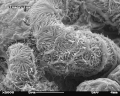
Glomerulus in the SEM. Image width 23 µm.
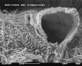
Glomerulus with broken capillary in the SEM. Image width 11.5 µm.

Internal view of the fenestrated endothelium ( fenestrae ) in the glomerulus of the mouse kidney in the SEM. Image width 1.15 µm.


Mesangium
The mesangium is a special type of connective tissue inside and outside the kidney corpuscle. The so-called mesangium cells ( mesangiocytes ) support the capillary walls, phagocytize and are also involved in the transmission of information during regulatory processes (tubuloglomerular feedback). The extra-glomerular mesangial cells are part of the juxtaglomerular apparatus .
function
In humans, about 1 liter of blood or 600 ml of blood plasma pass the glomeruli of the kidneys every minute ( renal plasma flow ), of which about 20%, i.e. about 120 ml per minute, are filtered ( glomerular filtration rate ). Around 180 liters of primary urine are produced per day. 80 to 90% of this is reabsorbed in the proximal tubules. The hormone-regulated fine - tuning takes place depending on ADH in the main cells of the collecting pipes , so that a total of 99% of the water is reabsorbed and around 1.5 liters of urine are formed per day.
The decisive factor for the filtration is the pressure difference, i.e. the difference between the various pressures in the capillaries and in the Bowman capsules, which are each made up of the hydrostatic and the colloid osmotic pressure . During the passage through the glomerulus, the hydrostatic pressure practically does not decrease, because the resistance is low due to the large overall cross-section of the capillaries connected in parallel. Since an ultrafiltrate is pressed out and the plasma proteins remain, the protein concentration and thus the colloid osmotic pressure rise continuously during the capillary passage, so that the effective filtration pressure drops and reaches zero at the end when the filtration equilibrium is reached.
embryology
In embryology , the terms Bowman's capsule, nephron, glomerulus and renal tubules are used synonymously for completely different structures with completely different functions in the first month of development. The two post-kidneys only begin to function in the second half of pregnancy . The urine is then excreted into the amniotic cavity . The amniotic fluid ( amniotic fluid ) is swallowed by the fetus and absorbed in its gastrointestinal tract . The urinary substances get into the mother's blood via the child's bloodstream and the placenta . Then they are excreted through the mother's kidneys and bladder .
See also
literature
- Uwe Gille: urinary and sexual system, urogenital apparatus. In: FV. Salomon, H. Geyer, U. Gille (ed.): Anatomy for veterinary medicine . 2nd ext. Edition. Enke-Verlag, Stuttgart 2008, ISBN 978-3-8304-1075-1 .
- Werner Linß, Jochen Fanghänel: Histology: cytology, general histology, microscopic anatomy . Walter de Gruyter, 1998, ISBN 3-11-014032-2 , pp. 207-209.
Individual evidence
- ^ Renate Lüllmann-Rauch, Friedrich Paulsen: Pocket textbook histology. 4th edition. Thieme Verlag, 2012, ISBN 978-3-13-151664-0 , p.466ff.
- ↑ Robert F. Schmidt, Florian Lang: Physiology of humans: With pathophysiology. 30th edition. Springer, 2007, ISBN 978-3-540-32908-4 , p. 688.
- ↑ a b Ursula Baum: Anatomy and Physiology. 7th edition. Volume 1, Elsevier, Urban & Fischer-Verlag, 2004, ISBN 3-930192-62-4 , p. 164.
- ^ Jan Langman : Medical Embryology , 5th edition, Thieme-Verlag, Stuttgart 1977, ISBN 3-13-446605-8 , pages 168 to 176.
Web links
- - Blood-urinary barrier at the University of Freiburg, Department of Anatomy