Nephron
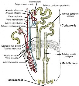
A nephron (from ancient Greek νεφρός nephros , German 'kidney' ) is the functional subunit of the kidney . The nephron is considered to be the smallest functional unit of the kidneys . It consists of:
- the renal corpuscle (Malpighi corpuscle, named after Marcello Malpighi ; corpusculum renale , with the glomerulum in Bowman's capsule ) and
- the connected kidney tubule ( tubule ).
Each human kidney has around a million of these subunits. The tubules work (despite a postulated tubuloglomerular feedback ) largely independently of the glomeruli.
physiology
Primary urine is continuously filtered from the blood in the kidney corpuscles . Then certain substances are resorbed in the tubules (primarily water is " reabsorbed ", reabsorbed , water reabsorption), but also secreted . This concentration creates the actual urine ( secondary urine or terminal urine ) from the primary urine . Primary urine formation is also called glomerular filtration , filtrative kidney function or creatinine clearance ; it is around 150 liters per day (or 105 ml / min) in adults.
The tubules regulate the water balance , the glomeruli filter the plasma in proportion to the variable cardiac output .
The actual kidney function consists in the active transport of the tubules (using energy) in contrast to the hemodynamically generated (passive) filtration of the glomeruli. The active transport processes in the kidney tubules are divided into primarily active, secondary active and tertiary active.
history
The theories of urination have a long history. Leonhart Fuchs (1501–1566) already described the kidney as a sieve or filter. The Austrian anatomist Josef Hyrtl also called a kidney a sieve (seyhe) or sieve . William Bowman still claimed in 1842 that the glomerular capillaries secrete water, which washes away the substances secreted by the tubules. Carl Ludwig also had a clear idea of how the kidneys work during urine preparation in 1842 . According to his mechanical theory , which is still essentially valid today , the physical filtration of the plasma takes place in the glomeruli. Then there is a back diffusion of water through an endosmosis in the tubule. The tubular resorption of urinary substances was not recognized until 1917 by Arthur Robertson Cushny . Today we speak of ( passive , i.e. without energy consumption) glomerular filtration and ( active , i.e. with energy consumption) tubular reabsorption . Franz Volhard already rejected this "modern mechanical-physical filtration theory", although he correctly described it in detail several times ("Filtration-re-absorption theory by Ludwig and Cushny"). The ( neurohumorally regulated and medically modulated) interplay of physics and chemistry in the podocytes and in the individual tubular sections in relation to the individual urinary substances has not yet been conclusively clarified.
Note
Only the tubules and their function are described below. The glomeruli , on the other hand, are dealt with under the heading kidney corpuscles . The unit of glomeruli and tubules is called the nephron. In the foreign-language Wikipedia encyclopedias, on the other hand, glomeruli and tubules can be found on an equal footing with the keyword nephron .
Overview of the tubular system
The renal tubule is divided into the main section ( proximal tubule), transition section (intermediate tubule or tubule attenuatus) and middle section (distal tubule). The straight sections of the kidney tubules and the connecting piece form a loop called Henle's loop (after Jakob Henle , Latin Ansa nephroni ). Henle's loops only exist in mammals and birds . They are obviously necessary to form urine that is hyperosmotic to the blood, because vertebrates without Henle's loops are not able to do so.
The connecting tubule and collecting tube are of a different embryological origin and therefore do not belong to the nephron. But they form a functional unit with the tubular system of the nephron. The distal tubule is distal with respect to the nephron.
In the nomenclature of the tubular system, anatomical and physiological aspects can be taken into account, which leads to different but complementary classifications.
Both the proximal and the distal tubule are divided into a "coiled" part, pars convoluta or pars contorta , and a "straight" part, pars recta . The rectal parts of both tubules and the intermediate tubule are functionally combined to form a loop of Henle . The pars recta of the distal tubule is often only referred to as the thick ascending part of the loop of Henle , while then under the distal tubule only the pars convoluta (also referred to as early distal tubule ) or even (referred to as late distal tubule ) the connecting tubule and the beginning of the Collecting tube are understood. The assignment of the connecting tubule to the middle piece or collecting tube is inconsistent. Here it is assigned to the manifold.
The following table compares German names, the names according to the noun anatomica , further classifications, international abbreviations and the anatomical position.
| Anatomical name | Other names | International | Anatomical location | physiology | histology | ||
|---|---|---|---|---|---|---|---|
| Main piece | Proximal tubule, pars convoluta | Proximal bundle | Proximal Convoluted Tubule (PCT) | bark |
Resorption of large amounts u. a. of Na + , glucose , bicarbonate and amino acids through Na + coupled symporters (glucose) or antiporters (bicarbonate)
Absorption or secretion, etc. a. of uric acid through anion transporters with the help of the proximal tubular cells |
high brush border, clear lumen, high density of mitochondria | |
| Proximal tubule, pars recta | Henle loop | Proximal Straight Tubule (PST) | Superficial nephrons: medullary rays
Middle nephrons: medullary rays, outer stripe of the outer marrow Juxtamedullary nephrons: outer stripe of the outer marrow |
||||
| Transition piece | Intermediate tubule, descending part | Descending thin part (thigh) of the loop of Henle, Pars descendens tubulus attenuatus |
Descending Thin Limb (DTL) | Superficial nephrons: medullary rays
Juxtamedullary and middle nephrons: inner stripe, outer marrow, inner marrow |
Concentration of urine using the countercurrent principle | flat epithelium | |
| Intermediate tubule, ascending pars | Ascending thin part (thigh) of the loop of Henle, Pars ascendens tubulus attenuatus |
Ascending Thin Limb (ATL) | Inner marrow, only present in juxtamedullary nephrons | Concentration of urine using the countercurrent principle | |||
| Middle piece | Distal tubule, pars recta | Thick ascending part (thigh) of the loop of Henle | Thick Ascending Limb (TAL) | Superficial nephrons: medullary rays, transition from the cortex
Juxtamedullary and middle nephrons: outer medulla, transition cortex |
Concentration of urine using the countercurrent principle | cubic, uniform epithelium, round cell nuclei, large mitochondria | |
| Distal tubule, pars convoluta | Distal convolute, early distal tubule |
Distal nephron | Distal Convoluted Tubule (DCT) | bark |
Aldosterone -dependent urine concentration, contains the macula densa |
||
| Manifold | Connecting tubule | late distal tubule, tubule reuniens | Connecting Tubule (CNT) | Bark, transition to medullary rays | Concentration of the urine by dehydration, ADH- dependent | cubic to prismatic cells, switching cells and main cells, heterogeneous, large lumen | |
| Manifold | Collecting Duct (CD) | Beginning at the top in medullary rays, runs through the whole medulla to the papilla | Concentration of urine through dehydration, ADH-dependent | ||||
Main piece

The main part ( tubulus proximalis ) runs first in a meandering manner ( tubulus contortus proximalis ) and then straight ( tubulus rectus proximalis ) into the renal medulla .
This is where the valuable compounds (e.g. glucose , amino acids , electrolytes ) contained in the primary urine are recovered. In addition, some pollutants are actively released here.
Transition piece
The transition piece ( tubulus attenuatus ) first pulls towards the renal medulla and then bends back towards the cortex. Here, the urine is mainly removed from water.
Middle piece
The middle section ( distal tubule ) begins in the renal medulla and initially extends into the renal cortex as a straight tube ( distal rectus tubule ). Here, in turn, there is a winding section ( tubulus contortus distalis ), which opens into a collecting tube.
In the distal tubule, NaCl is withdrawn from the urine and released into the renal medulla, where the NaCl returns to the bloodstream via the capillaries . Here an active transport takes place via ion channels : Na + is actively transported out, Cl - migrates passively.
Function of the tubules
The main task of the tubules is to reabsorb almost all of the primary urine into the bloodstream . In this respect, the tubular function is to be understood as the difference between primary urine and secondary urine. This subtraction applies not only to water, but also to all dissolved substances that are subject to urination. The tubular reabsorption of water ( volume per unit of time ) is equal to the difference between GFR and urine flow for any period . Likewise, the mass of the substances excreted in the urine is equal to the difference between the filtered and absorbed mass of the substance in question.
These differences are only falsified by the new formation of urinary substances in the tubules and by the tubular secretion of urinary substances in the secondary urine (mostly only very slightly). However, these two mechanisms are regularly negligible.
So oliguria and anuria are not indications of pathological disorders of glomeruli or tubules. In contrast, polyuria could be a symptom of diuretic therapy, polydipsia, or a rare tubular disease .
The more intense the tubular reabsorption, the less precise the determination of the glomerular filtration rate and creatinine clearance becomes . This is because all increases in the tubular reabsorption rate change the concentrations of urinary substances in the blood and urine.
Tubular diseases
All diuretics reduce the tubular resorption rate (tubular blockade ) and thus increase the secondary urine flow, resulting in polyuria. With polyuria, nocturia and polydipsia can also be expected. Tubular diseases ( tubular atrophy, tubulitis , tubular necrosis, tubulorhexis , tubulopathies, tubulonephrosis , "severe lesions of the tubular epithelia", re- absorption damage ) would reduce the active transport during re-absorption and therefore act like diuretics.
Actual tubulopathies with a diuretic effect are very rare. Examples are (nephrogenic) diabetes insipidus renalis as a special case of diabetes insipidus and the renal Fanconi syndrome . In hereditary Hartnup disease , Lowe syndrome and Gitelman syndrome , however, polyuria does not occur.
Isolated tubular diseases with an increased rate of re- absorption and consequently with the symptom of anuria are even rarer . Here is Liddle syndrome is an example. One speaks here of a pathological improvement in function (gain of function).
The tubulointerstitial nephritis is pronounced only in rare cases, so that there is a polyuria. "On the other hand, toxic pure tubular damage without a simultaneous impairment of kidney blood flow does not lead to insufficiency or anuria." Even with tubulointerstitial cell damage in the context of chronic renal insufficiency , polyuria does not occur. An acute tubular necrosis (obsolete term) as undesirable medication side effects not regularly leads to polyuria.
literature
- Karl Julius Ullrich , Klaus Hierholzer : Normal and pathological functions of the kidney tubule. Publisher Hans Huber, Bern / Stuttgart 1965, DNB 458762938 .
- John W. Boylan, Peter Deetjen, Kurt Kramer : Kidney and water balance. Urban & Schwarzenberg , Munich / Berlin / Vienna 1970, ISBN 3-541-04911-1 .
- Ulrich Kuhlmann u. a. (Ed.): Nephrology . 6th edition. Georg Thieme Verlag, Stuttgart / New York 2015, ISBN 978-3-13-700206-2 .
See also
Web links
Individual evidence
- ^ Robert Franz Schmidt , Florian Lang, Manfred Heckmann: Human physiology: with pathophysiology; with 85 tables; with removable revision course . 31., revised. and updated edition. Springer-Medizin-Verlag, Heidelberg 2010, ISBN 978-3-642-01650-9 .
- ↑ Karl Julius Ullrich , Klaus Hierholzer (Ed.): Normal and pathological functions of the kidney tubule. Verlag Hans Huber, Bern 1965, 466 pages.
- ↑ KH Gertz: The adaptation of transtubular absorption to the glomerular filtration rate. In: Karl Julius Ullrich , Klaus Hierholzer (eds.): Normal and pathological functions of the kidney tubule. Verlag Hans Huber, Bern 1965, pp. 141–145.
- ^ Heinz Valtin: Function of the kidney. 1st edition, Schattauer Verlag, Stuttgart / New York 1978, ISBN 3-7945-0556-5 , p. 35; Quote: "The absorption must be at least partially active".
- ↑ Another view ("The tubular reabsorption is a passive mechanism that affects the entire length of the tubule."), Albeit without justification, are Markus Daschner and P. Cochat: Pharmacotherapy for renal insufficiency. In: Karl Schärer, Otto Mehls (Ed.): Pediatric Nephrology. Springer-Verlag, Berlin, Heidelberg 2002, ISBN 978-3-642-62621-0 , p. 467.
- ^ EP Leumann: Kidney function tests. In: Karl Schärer, Otto Mehls (Ed.): Pediatric Nephrology. Springer-Verlag, Berlin, Heidelberg 2002, ISBN 978-3-642-62621-0 , p. 22.
- ^ Claas Wesseler: Physiologie , Volume 1, 3rd edition, Medi-Learn, Marburg 2009, ISBN 978-3-938802-58-8 , pp. 3–7.
- ↑ So the chapter heading in the table of contents on page 1 in: Franz Volhard : Die double-sided haematogenous kidney diseases . In: Gustav von Bergmann , Rudolf Staehelin (Hrsg.): Handbuch der Innere Medizin , 2nd edition, published by Julius Springer, Berlin / Heidelberg 1931, volume 6, first part, pages V and 1.
- ↑ Johanna Bleker : The history of kidney diseases. Boehringer Mannheim 1972.
- ^ Heinz Valtin: Function of the kidney. 1st edition, Schattauer Verlag, Stuttgart / New York 1978, ISBN 3-7945-0556-5 , p. 6.
- ^ William Bowman : On the structure and use of the malpighian bodies of the kidney, and observations on the circulation through that gland. Philosophical Transactions of the Royal Society , London, 132: p. 57 (1842).
- ^ Carl Ludwig : Kidneys and urine preparation. In: Rudolf Wagner (Hrsg.): Concise dictionary of physiology with consideration for physiological pathology. Vieweg, Braunschweig 1844.
- ↑ H. Straub, K. Beckmann: General pathology of water and salt metabolism and urine preparation. In: Textbook of Internal Medicine. 4th edition, 2nd volume, published by Julius Springer, Berlin 1939, p. 8.
- ↑ Arthur Robertson Cushny : The Secretion of Urine. Longmans, Green and Company, London 1917.
- ^ W. Kaiser: The years of ordination in Halle by Franz Volhard (1872-1950). In: Hans Erhard Bock , Karl-Heinz Hildebrand, Hans Joachim Sarre (eds.): Franz Volhard - memories. Schattauer Verlag, Stuttgart 1982, ISBN 3-7845-0898-X , p. 212.
- ↑ Franz Volhard : The bilateral hematogenous kidney diseases . In: Gustav von Bergmann , Rudolf Staehelin (Hrsg.): Handbuch der Innere Medizin , 2nd edition, published by Julius Springer, Berlin / Heidelberg 1931, Volume 6, first part, pages 18 and 21.
- ^ Heinz Valtin: Function of the kidney. 1st edition, Schattauer Verlag, Stuttgart / New York 1978, ISBN 3-7945-0556-5 , p. 36.
- ^ Karl Klütsch, Ernst Wollheim, Hans-Jürgen Holtmeier (eds.): The kidney in circulation , Georg Thieme Verlag, Stuttgart 1971, ISBN 3-13-468201-X , p. 148.
- ↑ Siegfried Waldeggerhaus, Martin Konrad: Tubular diseases (tubulopathies). In: Jörg Dötsch, Lutz T. Weber (Hrsg.): Kidney diseases in childhood and adolescence. Springer-Verlag, Berlin 2017, ISBN 978-3-662-48788-4 , p. 133.
- ^ Ulrich Kunzendorf: Kidney Transplantation. In: Ulrich Kuhlmann, Joachim Böhler, Friedrich C. Luft , Mark Dominik Alscher , Ulrich Kunzendorf (eds.): Nephrology . 6th edition. Georg Thieme Verlag, Stuttgart, New York 2015, ISBN 978-3-13-700206-2 , p. 776.
- ↑ The term acute tubular necrosis is now considered obsolete. See acute kidney failure .
- ↑ Hans Joachim Sarre : Kidney Diseases. 4th edition, Georg Thieme Verlag, Stuttgart 1976, ISBN 3-13-392804-X , p. 428.
- ↑ Karl Schärer, M. Konrad, W. Rascher, G. Reusz, Otto Mehls: Hereditary Tubulopathien. In: Karl Schärer, Otto Mehls (Ed.): Pediatric Nephrology. Springer-Verlag, Berlin, Heidelberg 2002, ISBN 978-3-642-62621-0 , pp. 119-148.
- ↑ O. Spühler: The interstitial nephritis and the importance of Franz Volhard for their teaching. In: Hans Erhard Bock , Karl-Heinz Hildebrand, Hans Joachim Sarre (eds.): Franz Volhard - memories. Schattauer Verlag, Stuttgart 1982, ISBN 3-7845-0898-X , p. 169.
- ↑ Franz Volhard : The bilateral hematogenous kidney diseases . In: Gustav von Bergmann , Rudolf Staehelin (Ed.): Handbook of internal medicine. 2nd edition, Springer-Verlag, Berlin, Heidelberg 1931, Volume 6, ISBN 978-3-662-42701-9 (reprint), p. 270.
- ↑ Hans Joachim Sarre : Kidney Diseases. 4th edition, Georg Thieme Verlag, Stuttgart 1976, ISBN 3-13-392804-X , p. 532 f.
- ^ Claas Wesseler: Physiologie , Volume 1, 3rd edition, Medi-Learn, Marburg 2009, ISBN 978-3-938802-58-8 , p. 32.
- ↑ O. Spühler: The interstitial nephritis and the importance of Franz Volhard for their teaching. In: Hans Erhard Bock , Karl-Heinz Hildebrand, Hans Joachim Sarre (eds.): Franz Volhard - memories. Schattauer Verlag, Stuttgart 1982, ISBN 3-7845-0898-X , p. 169.
- ↑ Otto Mehls, Karl Schärer: Chronic renal insufficiency. In: Karl Schärer, Otto Mehls (Ed.): Pediatric Nephrology. Springer-Verlag, Berlin, Heidelberg 2002, ISBN 978-3-642-62621-0 , p. 375.
- ↑ This term is now considered out of date. See acute kidney failure .
- ↑ P. Cochat, Markus Daschner: Nephrotoxicity of drugs. In: Karl Schärer, Otto Mehls (Ed.): Pediatric Nephrology. Springer-Verlag, Berlin, Heidelberg 2002, ISBN 978-3-642-62621-0 , p. 484.
- ^ C. Machleidt, Ulrich Kuhlmann: Interstitial nephropathies. In: Ulrich Kuhlmann, Joachim Böhler, Friedrich C. Luft , Mark Dominik Alscher , Ulrich Kunzendorf (eds.): Nephrology . 6th edition. Georg Thieme Verlag, Stuttgart, New York 2015, ISBN 978-3-13-700206-2 , pp. 512-515.

