
Wright stain
The Wright stain is a histological staining for displaying and differentiation of different blood cells .
properties
Wright stain is mainly used for blood smears and bone marrow biopsies. The cell nuclei are stained. Included dyes are Methylene Blue and Eosin Y or Eosin B . The methylene blue stains nucleic acids such as B. chromosomes , which is why Wright stain is also used for cytodiagnostics . The eosin stains basic areas within the cells such as B. DNA binding proteins .
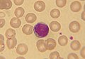
Wright stained lymphocyte

Basophil granulocyte stained according to Wright
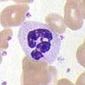
Wright stained neutrophil granulocyte


Variants of Wright stain are the buffered Wright stain, the Wright- Giemsa stain and the buffered Wright-Giemsa stain. An alternative to Wright staining is May-Grünwald staining and Pappenheim staining . The faster Wright stain is more common in the English-speaking world, while in Europe the Pappenheim stain and the Hemacolor stain are more commonly used.
history
Wright stain was developed by James Homer Wright in 1902 . It is a variant of the Romanowsky coloring .
Individual evidence
- ↑ Bernadette F. Rodak: Hematology. Elsevier Health Sciences, 2013, ISBN 978-0-323-29269-6 , pp. 197-198.
- ↑ a b Rolf Mahlberg: Hematology. John Wiley & Sons, 2014, ISBN 978-3-527-68125-9 , p. 221.
- ^ RE Lee, RH Young, B. Castleman: James Homer Wright: a biography of the enigmatic creator of the Wright stain on the occasion of its centennial. In: The American journal of surgical pathology. Volume 26, Number 1, January 2002, pp. 88-96, PMID 11756774 .