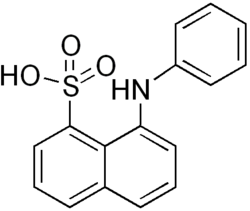
8-anilinonaphthalene-1-sulfonic acid
| Structural formula | ||||||||||||||||||||||
|---|---|---|---|---|---|---|---|---|---|---|---|---|---|---|---|---|---|---|---|---|---|---|

|
||||||||||||||||||||||
| General | ||||||||||||||||||||||
| Surname | 8-anilinonaphthalene-1-sulfonic acid | |||||||||||||||||||||
| other names |
|
|||||||||||||||||||||
| Molecular formula | C 16 H 13 NO 3 S | |||||||||||||||||||||
| Brief description |
gray powder |
|||||||||||||||||||||
| External identifiers / databases | ||||||||||||||||||||||
|
||||||||||||||||||||||
| properties | ||||||||||||||||||||||
| Molar mass | 299.34 g mol −1 | |||||||||||||||||||||
| Physical state |
firmly |
|||||||||||||||||||||
| Melting point |
215-217 ° C |
|||||||||||||||||||||
| safety instructions | ||||||||||||||||||||||
|
||||||||||||||||||||||
| As far as possible and customary, SI units are used. Unless otherwise noted, the data given apply to standard conditions . | ||||||||||||||||||||||
8-anilinonaphthalene-1-sulfonic acid (ANS) is a chemical compound from the group of sulfonic acids . ANS is used for fluorescent labeling of proteins . The light intensity increases when ANS binds to a hydrophobic region of a protein. ANS is used to stain the membranes of mitochondria . When proteins are denatured with chaotropes , the number of bound ANS molecules increases with unfolding .
Individual evidence
- ^ F. Mayer: Chemistry of organic dyes. Рипол Классик, ISBN 978-5-877-06767-7 , p. 55 ( limited preview in Google Book Search).
- ↑ a b c Data sheet 8-Anilino-1-naphthalenesulfonic acid from Sigma-Aldrich , accessed on May 22, 2017 ( PDF ).
- ↑ A. Málnási-Csizmadia, G. Hegyi, F. Tölgyesi, AG Szent-Györgyi, L. Nyitray: Fluorescence measurements detect changes in scallop myosin regulatory domain. In: FEBS Journal . Volume 261, Number 2, April 1999, pp. 452-458, PMID 10215856 .
- ↑ N. Gains, AP Dawson: 8-Anilinonaphthalene-1-sulphonate interaction with whole and disrupted mitochondria: a re-evaluation of the use of double-reciprocal plots in the derivation of binding parameters for fluorescent probes binding to mitochondrial membranes. In: Biochem J . Volume 148, Number 1, April 1975, pp. 157-160, PMID 1156395 , PMC 1165518 (free full text).
- ↑ VN Uversky, S. Winter, G. Löber: Use of fluorescence decay times of 8-ANS-protein complexes to study the conformational transitions in proteins which unfold through the molten globule state. In: Biophysical Chemistry . Volume 60, Number 3, June 1996, pp. 79-88, PMID 8679928 .
