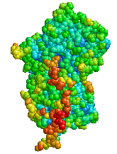
Alpha-1 antitrypsin deficiency
| Classification according to ICD-10 | |
|---|---|
| E88.0 | Plasma protein metabolism disorders, not elsewhere classified |
| ICD-10 online (WHO version 2019) | |
The α 1 -antitrypsin deficiency (synonyms: Laurell-Eriksson syndrome , protease inhibitor deficiency , AAT deficit ) is a hereditary metabolic disease due to a polymorphism of the proteinase system. A deficiency in protease inhibitors can lead to cirrhosis of the liver and emphysema .
history
Antitrypsin mutations may have spread throughout Europe during the Iron Age as they provided an evolutionary advantage in focusing and amplifying the inflammatory response to limit invasive gastrointestinal and respiratory infections. Only since the discovery of antibiotics have these protective mutations become harmful due to the spread of smoking and the longer lifespan.
Carl-Bertil Laurell and Sten Eriksson were the first to describe the disease in the early 1960s. There was an increased incidence of emphysema in one family.
genetics
- Inheritance: autosomal - co-dominant (AR)
- Gene locus: chromosome 14q32.1
- Frequency: 1: 2000 to 1: 5000, the disease is often not recognized
- Mechanism: Unhindered proteolysis of the tissue by proteases (mainly from neutrophils)
Mutations at position 342 (PiZZ) always lead to clinical symptoms, but mutations at position 264 (PiSS) usually remain silent.
| Lifetime risk by genotype | ||||
|---|---|---|---|---|
| genotype |
Frequency in Europe |
Typical serum concentration |
Emphysema - risk |
Cirrhosis - risk |
|
PiMM (normal) |
91.1% | 20-53 µmol / L 102-254 mg / dL |
normal | normal |
|
PiMS (carrier, subclinical) |
6.6% | 18-52 µmol / L
86-218 mg / dL |
normal | normal |
|
PiMZ (carrier, mild to moderate) |
1.9% | 15-42 µmol / L
62-151 mg / dL |
slightly increased | possible |
|
PiSS (homozygous, mild) |
0.3% | 20-48 µmol / L
43-154 mg / dL |
slightly increased | normal |
|
PiSZ (moderate) |
0.1% | 10-23 µmol / L
33-108 mg / dL |
slightly increased
to increased * |
possible |
|
PiZZ (homozygous, difficult) |
0.01% | 3.4-7 µmol / L
<29-52 mg / dL |
high | high |
| Pi00 | - | 0 μmol / L
0 mg / dL |
very high | normal |
- * Based on empirical data, it has been determined that a serum concentration <57 mg / dL is associated with an increased risk of lung disease.
Pathogenesis
The α 1 -antitrypsin is an acute phase protein and one of the most important proteinase inhibitors in serum . Among other things, it inhibits the proteinases trypsin and neutrophil elastase (NE). A deficiency can lead to increased proteolysis . The normal serum concentration is 0.9–2.0 g / l.

The mutations lead to a structural change in the proteins . This results in disturbed secretion and malfunction. This leads to aggregation and accumulation in the endoplasmic reticulum (ER) of the liver cells (hepatocytes) and subsequently to a deficiency in the cytoplasm (usually below 40% of normal). This results in decreased proteinase inhibitor activity and thus increased proteolysis.
The uninhibited leukocyte elastase destroys the lung structure. Progressive emphysema develops . The accumulation of α 1 -antitrypsin in the endoplasmic reticulum of the hepatocytes leads to cell damage and subsequently to fibrosis and liver cirrhosis .
Symptoms
Most patients have chronic, active liver inflammation ( hepatitis ), which is often noticeable in childhood. In later life, up to 40% of those affected develop cirrhosis of the liver and around 15% develop hepatocellular carcinoma ( HCC ).
The most important manifestation in homozygotes is chronic obstructive pulmonary disease (COPD). This usually only occurs after the age of 30. The emphysema and complications of COPD can use the respiratory failure to hypoxemia with possible right heart failure and pulmonary heart disease to multiple organ failure lead. In some cases the breathing pump can become exhausted and hypercapnia with increased arterial carbon dioxide partial pressure can be a complication.
Occasionally, glomerulonephritis , necrotizing vasculitis , granulomatosis with polyangiitis , necrotizing panniculitis , pancreatitis, and pancreatic fibrosis occur .
Diagnosis
The diagnosis is recommended once for all patients with COPD. This is especially true in the case of an unusual course, younger people and non-smokers who develop COPD.
The diagnostic criteria for the detection of an α 1 -antitrypsin deficiency are:
- α 1 -antitrypsin <0.9 g / l
- Proof of the genotypes PiZZ, PiMZ and PiSZ
- PAS-positive, protease-resistant hepatocellular inclusion bodies (= antitrypsin deposits)
It often takes several years from the first symptoms to diagnosis. Many patients are discovered too late, they are only advised to quit smoking, and they are treated inadequately and without proper screening for years.
therapy
In addition to the substitution that is required in some cases, the secondary diseases are primarily treated, especially chronic obstructive pulmonary disease . It is absolutely necessary to refrain from smoking completely , as the oxidants contained in the smoke inactivate α 1 -antitrypsin. It is also advisable to stay away from fire smoke and dust (hay, grinding work) of any kind or to wear respiratory protection. The latter is sometimes recommended to sensitive people in order to try to protect themselves from infections. Appropriate drug treatment is usually necessary to keep the consequences of COPD and especially infections as low as possible.
In this context, vaccinations are also recommended ( flu , pneumococci ).
Substitution therapy
In severe pulmonary emphysema , intravenous replacement ( substitution therapy ) of α 1 -antitrypsin is recommended in some cases . A level above 0.8 g / l should be aimed for in order to return to the range of clinically normal people. Substitution does not bring any advantage in the presence of liver damage , because the focus here is on accumulation .
Organ transplant
A lung or liver transplant may be necessary in the advanced stages . Liver transplantation is curative because α 1 -antitrypsin is hardly synthesized in extrahepatic tissue, but does not repair the lung damage that has already occurred.
Associated diseases
α 1 -antitrypsin deficiency is associated with a number of diseases:
- Cirrhosis of the liver
- COPD
- Pneumothorax
- asthma
- Granulomatosis with polyangiitis
- Pancreatitis
- Cholecystolithiasis (gallstones)
- Bronchiectasis
- Prolapse of pelvic organs
- Primary sclerosing cholangitis
- Autoimmune hepatitis
- Emphysema , predominantly affects the lower lobes and causes bullae
- Carcinoma ( cancer )
literature
- Alexander Biedermann, Thomas Köhnlein: Alpha-1-antitrypsin deficiency - a hidden cause of COPD: overview of pathogenesis, diagnostics, clinical features and therapy. Deutsches Ärzteblatt , Volume 103, Issue 26 of June 30, 2006, pp. A-1828 / B-1569 / C-1518
- L. Fregonese, J. Stolk: Hereditary alpha-1-antitrypsin deficiency and its clinical consequences. In: Orphanet J Rare Dis. 2008 Jun 19; 3, p. 16. Review. PMID 18565211 , PMC 2441617 (free full text)
- EK Silverman, RA Sandhaus: Clinical practice: Alpha1-antitrypsin deficiency. In: New England Journal of Medicine. (2009) vol 360 (26), pp. 2749-2757. Review. PMID 19553648
- J. Stoller, L. Aboussouan: Alpha1 -antitrypsin deficiency. In: Lancet. (2005) 365 (9478), pp. 2225-2236. PMID 15978931
Web links
Individual evidence
- ↑ David A. Lomas: The Selective Advantage of α-Antitrypsin Deficiency. In: American Journal of Respiratory and Critical Care Medicine. 173, 2006, p. 1072, doi : 10.1164 / rccm.200511-1797PP .
- ↑ Christoph Höner zu Siederdissen, Thomas Köhnlein: Alpha-1-Antitrypsinmangel . In: Practice of Hepatology . Springer Berlin Heidelberg, Berlin, Heidelberg 2016, ISBN 978-3-642-41620-0 , pp. 155-160 , doi : 10.1007 / 978-3-642-41620-0_24 .
- ↑ James K. Stoller, Felicitas L. Lacbawan, Loutfi S. Aboussouan: Alpha-1 Antitrypsin Deficiency . In: GeneReviews (®) . University of Washington, Seattle, Seattle (WA) January 1, 1993, PMID 20301692 ( nih.gov [accessed February 1, 2017]).
- ^ Sarah K. Brode, Simon C. Ling, Kenneth R. Chapman: Alpha-1 antitrypsin deficiency: a commonly overlooked cause of lung disease . In: CMAJ: Canadian Medical Association Journal . tape 184 , no. 12 , September 4, 2012, ISSN 0820-3946 , p. 1365-1371 , doi : 10.1503 / cmaj.111749 , PMID 22761482 , PMC 3447047 (free full text).
- ↑ Vogelmeier, C et al., Guideline COPD, Pneumology 2007; 61: e1-e40.
