Ophthalmoscopy
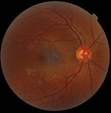
The ophthalmoscopy ( gr. Ὀφθαλμοσκοπία , ophthalmoskopia - "the perception of the eye") or ophthalmoscopy or fundoscopy (from the Latin fundus within the meaning of fundus ) is inspection to assess the visible parts of the eye . In particular, the retina and the blood vessels supplying it can be examined. The arteries that arise from the optic nerve papilla (blind spot) and appear light-red cross the dark-red veins of the retina.
An examination of the fundus with pupil-dilating medication is often prepared in order to provide a better insight into what is associated with the subject's temporary inability to drive.
history
The experiments to illuminate the fundus of the eye, carried out by Hermann von Helmholtz in 1850/51, based on Ernst Brücke in 1847 , developed an ophthalmoscope that illuminates the retina and clearly shows it (using a concave lens) Insight into the interior of an organ. Unlike an endoscope , it does not physically penetrate the organ. The ophthalmologist and medical historian Friedrich Hel Reich (1842–1927), who completed his habilitation in 1869 with a thesis on glaucoma and had operated a private eye clinic in Würzburg from 1872 , first asked for the inclusion of the ophthalmoscope in the basic equipment of clinics for internal medicine .
species
Ophthalmoscopy can be done in two different ways:

- In direct ophthalmoscopy , a concave mirror with a viewing hole or a converging lens in the middle for illuminating the fundus is used as a so-called direct ophthalmoscope and is brought very close between the patient's eye and the examiner's eye. The distance between examiner and patient is approx. 10 cm, so that the examination is often perceived as uncomfortable.
- In indirect ophthalmoscopy , an illuminated section of the fundus is viewed from a distance of approx. 50 cm using a light source and a magnifying glass held 2–10 cm in front of the patient's eye .
With direct ophthalmoscopy, the central parts such as the optic nerve head , vascular origins and the yellow spot (macula lutea) can be viewed easily and with the magnification brought about by the lens of the eye.
With indirect ophthalmoscopy, the retina , the optic nerve , the vessels, the macula lutea (yellow spot) and the retinal periphery can be easily examined. The magnification is not as strong as with direct ophthalmoscopy, but the overview is much better here and, in contrast to direct ophthalmoscopy, a stereoscopic (3D) assessment is possible, so that most ophthalmologists prefer this examination technique. In addition, indirect ophthalmoscopy can also be performed on the slit lamp . This enables the retinal image to be enlarged or assessed by projecting a light slit (even stronger 3D effect ). Another instrument for indirect reflection is the bonoscope .
In recent years, so-called scanning laser ophthalmoscopes have been brought to market maturity in the field of imaging processes , which can produce high-resolution three-dimensional slice or relief representations by means of point-by-point or line-by-line scanning of the retina and confocal aperture and lighting technology . Patients usually remain fit to drive by using this procedure, as they are not given any medication to dilate their pupils , and can view the images of their eyes for themselves. However, the application of the procedure is not covered by statutory health insurances ( individual health service ).
See also
Web links
- DIN NA 027 Standards Committee Precision Mechanics and Optics (NAFuO) - Ophthalmic Instruments - Direct Ophthalmoscopes, DIN EN ISO 10942.
- DIN NA 027 Standards Committee Precision Mechanics and Optics (NAFuO) - Ophthalmic Instruments - Indirect Ophthalmoscopes, DIN EN ISO 10943.
literature
- Holger Dietze, Antje Albaladejo Gomez: Ophthalmoscopy. DOZ-Verlag Optical Specialist Publication GmbH, Heidelberg 2013, ISBN 978-3-942873-16-1 .
- Theodor Axenfeld (founder), Hans Pau (ed.): Textbook and atlas of ophthalmology. With the collaboration of Rudolf Sachsenweger and others 12th, completely revised edition. Gustav Fischer, Stuttgart et al. 1980, ISBN 3-437-00255-4 .
-
The invention of ophthalmoscopy, represented in the original descriptions of the ophthalmoscope by Helmholtz, Ruete and Giraud-Teulon. Introduced and explained by Wolfgang Jaeger, Brausdruck GmbH, Heidelberg 1977; enclosed the following reprints:
- H. Helmholtz: Description of an eye mirror for examining the retina in the living eye. A. Förstner'sche Verlagsbuchhandlung, Berlin 1851
- CG Theod. Ruete: The ophthalmoscope and the optometer for practical doctors. Dieterichsche Buchhandlung, Göttingen 1852
- Giraud-Teulon: Ophthalmoscopie binoculaire ou s'exerçant par le concours des deux yeux associés. J. Van Buggenhoudt, Brussels 1861
- W. Münchow: History of ophthalmology. Stuttgart 1984, pp. 576-594.
Individual evidence
- ↑ Jutta Schickore: Ophthalmoscope . In: Werner E. Gerabek , Bernhard D. Haage, Gundolf Keil , Wolfgang Wegner (eds.): Enzyklopädie Medizingeschichte. De Gruyter, Berlin / New York 2005, ISBN 3-11-015714-4 , p. 121.
- ↑ H. von Helmholtz: Description of an ophthalmoscope for examining the retina in the living eye. Berlin 1851.
- ^ Richard Toellner: Illustrated history of medicine. Andreas & Andreas, Salzburg 1990, Volume 3, p. 1202.
- ↑ Werner E. Gerabek: Hel Reich, Friedrich Wilhelm von. In: Werner E. Gerabek et al. (Ed.): Enzyklopädie Medizingeschichte. 2005, p. 565.
- ^ Albert J. Augustin: Ophthalmology. 3rd, completely revised and expanded edition. Springer, Berlin et al. 2007, ISBN 978-3-540-30454-8 , p. 961 ff.
- ↑ retinal exam , auge-online.de, accessed November 23, 2013.
