Charcot foot
| Classification according to ICD-10 | |
|---|---|
| M14.6 * | Neuropathic Arthropathy |
| ICD-10 online (WHO version 2019) | |
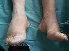
The Charcot foot is a disease pain sensitive feet, breaking unnoticed in bone without those affected feel fracture pain. 95% percent of all patients are diabetics . The incidence of acute illness in diabetics is between 0.15 and 2.5%. Charcot's foot is one of several diseases that are grouped under the umbrella term diabetic foot syndrome . The disease is named after the neurologist Jean-Martin Charcot . Another term that indicates the cause of the condition is neuroarthropathy . Neuroarthropathy of the foot was first described by the English doctor Herbert William Page in 1881. The term Charcot's foot was first used by Ralph H. Major in the American journal JAMA on May 17, 1928 on p. 846.
Causes of Neuroarthropathy
- Diabetic polyneuropathy
- syphilis
- leprosy
- Syringomyelia
- Meningomyeoloceles
- congenital neuropathies with impaired pain perception
- acquired neuropathies with disturbance of pain perception, e.g. B. Long-term alcohol abuse
Development of the disease
To date, the origin has not been precisely clarified. The prerequisite is the loss of pain sensitivity in the feet. The clinical picture is triggered by a skeletal injury (trauma). The neurovascular theory describes an increased blood flow and increased bone resorption caused by a nervous malfunction. The neurotraumatic theory states that if there is a lack of pain perception due to excessive strain, repeated minimal injuries to the joint surfaces occur, which then lead to increasing destruction of the bone, since this increasing damage is also perceived as hardly or not painful. Since there is no usual pain sensation, those affected continue to put strain on the broken foot and only consult a doctor if swelling, redness and deformity are very pronounced, but pain occurs or bone fragments are also painlessly pierced through the skin.
Diagnosis

- Anamnesis : As far as you can remember, the triggering trauma is often a banal injury, e. B. a sprain (distortion). If pain is indicated, it contradicts the examiner's own understanding of pain.
- Inspection : In the early active stage, the foot is partially or completely inflamed and reddened, without an infection (e.g. a rose or a phlegmon). Both feet are rarely affected at the same time. In the advanced active stage, the foot is hot, red, swollen and deformed, sometimes with open wounds on protruding bones. In the inactive, i.e. H. After the healed stage, there are no longer any clinical signs of inflammation such as reddening, swelling and overheating - any skeletal deformities persist.
- Examination: Skin dry, lack of sweat secretion (and lack of sweat odor), hyperkeratosis , overheated (temperature difference to the opposite side> 1 ° C), movement of the foot is painless in all joints despite the sometimes drastic deformities, open wounds, even if pus-filled, can be painless examined with surgical instruments. The other foot often appears completely unremarkable, but also shows complete numbness. In rare cases, however, both feet can be affected.
- Quantitative sensory testing (QST): the perception thresholds for thermal and mechanical pain stimuli ( pain thresholds ) are increased
- Imaging: X-ray diagnostics , computed tomography , magnetic resonance imaging / magnetic resonance imaging, possibly a leukocyte scintigraphy to rule out any suspicion of osteomyelitis . Diagnostics based solely on conventional X-ray images is nowadays viewed by the Cologne Higher Regional Court as inadequate.
Classification
The assessment of the Charcot foot is based on the one hand on the clinical findings and the radiologically visible course (classification according to Eichenholtz and Levin ), on the other hand according to the involvement of the bony structures (classification according to Sanders and Frykberg ).
| Stage of progress | Radiological signs | Clinical signs |
|---|---|---|
| 0 initial stage |
X-ray -native: normal MRI : bone marrow edema |
painless soft tissue swelling and reddening without evidence of infection |
| I stage of destruction Ia phase of fragmentation Ib phase of dislocation |
X-ray / MRI: demineralization osteolysis osseous fragmentation joint destruction |
Signs of inflammation (swelling, reddening, overheating). Joint deformation due to joint effusion. Joint instability. Increasing formation of foot deformity. Development mostly painless due to the polyneuropathy |
| II stage of reparation | Remineralization Fragment resorption New bone formation (formation of osteophytes, callus, sclerotherapy) |
Swelling, redness and overheating recede |
| III stage of consolidation | bony fusion with joint stiffening (ankylosis) | permanent foot deformity |
| IV stage of ulcer | deformed foot skeleton | Deformed foot with high risk of ulcer formation and risk of wound infection, foot is painless |
| Type | Infested structure | frequency |
|---|---|---|
| I. | Interphalangeal joints, metatarso-phalangeal joints, metatarsals | 10-30% |
| II | Tarso-metatarsal joints | 15-48% |
| III | Naviculo-cuneiform joints, talo-navicular joint, calcaneo-cuboid joint | 32% |
| IV | Ankle joints | 10% |
| V | Calcaneus | 2% |
therapy
- Acute Charcot's foot is an urgent emergency that requires immediate complete pressure relief and treatment in a specialized facility.
- First, complete immobilization (this means inpatient treatment at the beginning of diagnostics and therapy)
- After the acute phase has subsided, a plaster cast , a two-shell orthosis or a special rigid plastic bandage ( Total Contact Cast ) is adjusted until the bone destruction has completely healed, sometimes in a functional malposition.
- Then wearing a special orthopedic made-to-measure shoe
- In some cases, foot amputation may be necessary. In this case, a lower leg orthosis is made with which the orthotic shoe can be worn.
- Normalization of the sugar metabolism through adequate therapy for diabetes mellitus
- Training the patient
Monitoring the success of the treatment with magnetic resonance imaging (MRI)
When monitoring the healing process by means of MRI, it must be ensured that the fresh fracture callus in the first phase of the so-called secondary fracture healing runs under the MRI image of the " bone marrow edema " and can therefore hardly be distinguished from the "bone marrow edema" of the acute bone injury (at most by minor inflammatory swelling of the surrounding soft tissues). In the case of "bone marrow edema", a distinction must always be made as to whether it is a healing-related or an injury-related phenomenon. In the case of bone injuries that are limited to the microtrabeculae of the cancellous bone (without injuring the cortex), primary healing is more likely (without callus formation) and the "bone marrow edema" will gradually recede. If the cortex is injured , secondary healing with the formation of fracture callus and a discontinuous decrease (i.e. with a temporary increase) of the "bone marrow edema" can be expected. Increase in "bone marrow edema" can also be the result of renewed acute bone injury as a result of premature termination of the immobilization treatment.
Prominent patients
The French painter Édouard Manet (1832–1883) was a prominent patient . He suffered from incurable neurolues with ataxia since the late 1870s. After he twisted his left foot at the end of 1878 or beginning of 1879 and could no longer walk much, his doctor, Dr. Siredey 1880 climbing the stairs. He only limped, he had to lean on a stick, felt he was perceived as a "cripple". In the autumn of 1882, Manet wrote his will, because ulcers and bones had evidently formed and his general condition worsened. When the foot became 'gangrenous' and there was a risk of sepsis, the left leg was amputated on April 20, 1883. The painter died on April 30th as a result of the surgery.
literature
- Franz X. Koeck, Bernhard Koester: Diabetic foot syndrome. Thieme, Stuttgart 2007, ISBN 978-3-13-140821-1 . (Medical textbook)
- H. Reike: Diabetic foot syndrome: diagnosis and therapy of the underlying diseases and complications. de Gruyter, Berlin 1998, ISBN 3-11-016215-6 .
- Ludger Poll, Ernst Chantelau: Charcot foot: It depends on the early diagnosis . In: Deutsches Ärzteblatt . tape 107 , no. 7 , 2010, p. A-272 ( aerzteblatt.de [accessed October 13, 2016]).
Web links
- Evidence-based guideline: diagnosis, therapy, follow-up and prevention of diabetic foot syndrome . (PDF; 573 kB) German Diabetes Society, German Society for Vascular Surgery
- S3 guideline National care guideline for type 2 diabetes: prevention and treatment strategies for foot complications of the BÄK, KBV, AWMF. In: AWMF online (as of 2006)
Individual evidence
- ↑ Alphabetical index for the ICD-10-WHO version 2019, volume 3. German Institute for Medical Documentation and Information (DIMDI), Cologne, 2019, p. 149
- ↑ Charcot Arthropathy on emedicine.com
- ↑ Lee J. Sanders, Michael E. Edmonds, William J. Jeffcoate: Who was first to diagnose and report neuropathic arthropathy of the foot and ankle: Jean-Martin Charcot or Herbert William Page? In: Diabetologia . tape 56 , no. 9 , September 1, 2013, p. 1873-1877 , doi : 10.1007 / s00125-013-2961-6 .
- ↑ Ernst A. Chantelau, Gotthard Grützner: Is the Eichenholtz classification still valid for the diabetic Charcot foot? Swiss Medical Weekly, smw.ch, April 24, 2014, accessed on March 29, 2016 .
- ↑ Ernst A. Chantelau: Nociception at the diabetic foot, an uncharted territory . In: World Journal of Diabetes . tape 6 , 2015, p. 391-402 ( wjgnet.com [accessed November 23, 2016]).
- ↑ R. Ramnarine, A. Isaac, LM Meacock, N. Petrova, M. Edmonds, DAElias. A novel semi-quantitative scoring proforma in the assessment of radiological resolution of the acute diabetic Charcot foot. http://posterng.netkey.at/essr/viewing/index.php?module=viewing_poster&task=viewsection&pi=131064&ti=443216&si=1508&searchkey=#poster
- ↑ Barbara Berner: Legal report. Liability of a doctor in attendance. In: Deutsches Ärzteblatt Vol. 116 issue April 15 , 2019, p. C 606 , accessed on May 15, 2019 .
- ↑ Joachim Grifka (Ed.): Diabetic foot syndrome. Verlag Georg Thieme, Stuttgart 2007, ISBN 978-3-13-140821-1 .
- ↑ nice.org.uk
- ↑ Wolf-Rüdiger Klare et al .: Total Contact Cast: Effective in the treatment of diabetic foot ulcers (PDF; 2.2 MB)
- ↑ deutsche-diabetes-gesellschaft.de ( Memento of the original from September 23, 2015 in the Internet Archive ) Info: The archive link was inserted automatically and has not yet been checked. Please check the original and archive link according to the instructions and then remove this notice. (PDF; 1.9 MB) DDG Practice Guideline for Diabetic Foot Syndrome 2010.
- ↑ Ernst-Adolf Chantelau, Sofia Antoniou, Brigitte Zweck, Patrick Haage: Follow up of MRI bone marrow edema in the treated diabetic Charcot foot - a review of patient charts. In: . Diabetic Foot & Ankle. 2018, accessed May 15, 2019 .
- ^ Réunion des Musées Nationaux Paris, Metropolitan Museum of Art New York (ed.): Manet. Exhibition catalog, German edition: Frölich and Kaufmann, Berlin 1984, ISBN 3-88725-092-3 .
- ^ Antonin Proust: Édouard Manet, memories. Berlin 1917

