
Fascia

|
This article was based on formal and / or substantive defects on the quality assurance side of the editorial Medicine entered. Please help fix the shortcomings in this article and join the discussion there . The minimum requirements for medical articles are to be met, thereby avoiding a possible deletion of the article or article passages within four weeks.
Reason: See WP: Redaktion_Medizin / Qualitätssicherung # Faszie, _Faszientraining |
 |


Fascia , and the fascia (borrowing from latin fascia , Band ' , bandage') denotes the soft components of the connective tissue , which penetrate the entire body as a covering and connecting voltage network. This includes all collagenous fibrous connective tissue, especially the joints and organ capsules , tendons, plates ( aponeuroses ) Muskelsepten, ligaments , tendons , retinacula (so-called "straps", for example, the carpal tunnel forming the flexor retinaculum ) and the "actual" fascia in the form of " Muscle ties ”such as B. the thoracolumbar fascia , which surrounds the erector spinae muscle like a stocking.
Some authors occasionally use a narrower fascia definition, according to which only flat structures are referred to as fascia. Depending on the author, aponeuroses, retinacula, the superficial fascia (subcutaneous fat tissue) or intramuscular connective tissue may or may not be included. Since the first international Fascia Research Congress in 2007, the leading experts in this field have agreed on the more comprehensive concept of fascia formulated above. This new definition of fascia is essentially congruent with what the layman understands by “ connective tissue ” (in contrast to the medical practitioner, for whom, for example, cartilage and bone tissue are also counted as connective and supporting tissue).
The three layers of the fascia
Superficial fascia
Superficial fasciae are found in the subcutaneous tissue in most parts of the body and mix with the reticular layer of the dermis . They are located above the upper area of the sternocleidomastoid muscle , on the neck and above the breastbone ( sternum ). They mainly consist of loose connective tissue and fat tissue. In addition to their subcutaneous presence, this type of fascia encloses organs, glands and neurovascular channels and fills free space in many other places. It stores fat and water; it acts as a passage for lymph , nerves and blood vessels as well as a buffer and damper. Since a considerable part of the connective tissue cells in this layer is in contact with one another, it is also assumed that this layer could serve as a body-wide non-neural communication network.
Deep fascia
Deep fasciae are the dense, fiber-rich connective tissue layers and strands that penetrate and enclose the muscles, bones, nerve tracts and blood vessels of the body. Depending on the local stress conditions, this tissue network condenses and organizes itself as tendon plates ( aponeuroses ), large flat fascia (such as the fascia lata or the plantar fascia ), as ligaments (ligaments), retinacula (fetters), joint capsules or as muscle septa. As a highly innervated periosteum, this tissue envelops the bones, as the perichondrium the cartilage tissue, as the tunica externa the blood vessels and as the perineurium the nerve tracts. In addition, all muscle fibers are covered by an endomysium layer, while the perimysium combines individual muscle fiber bundles and finally the epimysium covers the entire muscle. The high proportion of collagen fibers gives these fabrics a high viscoelastic tensile strength.
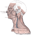
The galea aponeurotica and the temporal fascia

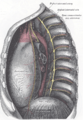
The fascia of the diaphragm

The aponeurosis of the obliquus externus abdominis muscle (external oblique abdominal muscle)

The deep ligaments of the arch of the foot


Visceral fascia
Visceral fascia are used to suspend and embed the internal organs and wrap them in layers of connective tissue membranes. Each of these organs is surrounded by a double layer of serous membranes. The outermost wall of an organ is called the "parietal layer" while the skin of the organ is called the "visceral layer". The organs have specific names for their visceral fascia. In the brain they are called meninges , in the heart the pericardium , in the lungs the pleura and in the abdomen the peritoneum .

The meninges
The pericardium and the left dome of the diaphragm

The peritoneum with the kidney fascia


Fascial dynamics
Fascia are very adaptable parts of tissue.
- Because of their high viscoelasticity , superficial fasciae can stretch significantly, for example to absorb body fat in connection with normal or prenatal weight gain.
- Visceral fasciae are generally less flexible than the superficial fasciae. Because of their connecting function for the organs, their tension must remain constant. If they were too loose it would lead to an incident of the organ; if they were too hypertonic , it would restrict organ mobility.
- Deep fasciae are also less flexible than superficial fasciae. They have less blood flow, but are highly innervated with sensory receptors that signal pain ( nociceptors ), changes in movement ( proprioceptors ), changes in pressure and vibrations ( mechanoreceptors ), changes in the chemical environment ( chemoreceptors ) and temperature fluctuations ( thermoreceptors ). Many deep fasciae are able to respond to appropriate mechanical or chemical stimulation with contraction or relaxation as well as through a gradual structural reorganization of their internal components. Deep fasciae have special smooth muscle- like connective tissue cells ( myofibroblasts ), which give them the ability to actively contract over a long period of time, like many intestines or blood vessels. The stiffness of a fascia is apparently related to the density of myofibroblasts. A particularly high density of myofibroblasts is found in both palmar fibromatosis ( Dupuytren's contracture ) and pathological frozen shoulder (“ frozen shoulder ”).
- Numerous manual therapeutic procedures aim to trigger a lasting change in the fascia. These include connective tissue massage , osteopathy , Rolfing , fascia therapy and cupping . Cupping is primarily aimed at changing the superficial fascia, while z. B. Rolfing and osteopathy intend to affect the deep fascia.
training
The fasciae can be specifically trained using various methods, e.g. B. using a fascia roller .
See also
literature
- Robert Schleip, Thomas W. Findley, Leon Chaitow, Peter A. Huijing (eds.): Textbook fascia - basics, research, treatment . Urban & Fischer Verlag / Elsevier, Munich 2014, ISBN 978-3-437-55306-6 .
- Carla Stecco: Atlas of the human fascia system. Urban & Fischer Verlag / Elsevier, 2016, ISBN 978-3-437-55905-1 .
Web links
Individual evidence
- ↑ fascia | German »Latin |. In: Pons. Retrieved February 24, 2017 .
- ↑ About Fascia. In: fasciacongress.org. Ida P. Rolf Research Foundation, accessed May 22, 2019 .
- ^ First International Fascia Research Congress. (English)
- ↑ John E. Skandalakis, PN Skandalakis, LJ Skandalakis, J. Skandalakis: Surgical Anatomy and Technique . 2nd Edition. Springer, Atlanta 2002, ISBN 0-387-98752-5 , pp. 1-2.
- ↑ Serge Paoletti: The Fasciae. Anatomy, Dysfunction & Treatment. Eastland Press, Seattle 2006, ISBN 0-939616-53-X , pp. 23-24.
- ↑ Gil. Hedley: The Integral Anatomy Series . Volume 1: Skin and Superficial fascia . DVD. Integral Anatomy Productions, 2005.
- ^ M. Langevin: Connective tissue: A bodywide signaling network? PMID 16483726 .
- ↑ Gil. Hedley: The Integral Anatomy Series . Volume 2: Deep Fascia and Muscle . DVD. Integral Anatomy Productions, 2005.
- ↑ Gil. Hedley: The Integral Anatomy Series . Volume 3: Cranial and Visceral Fasciae . DVD. Integral Anatomy Productions, 2005.
- ↑ Serge Paoletti: The Fasciae. Anatomy, Dysfunction & Treatment . Eastland Press, Seattle 2006, ISBN 0-939616-53-X , pp. 146-147.
- ^ Ida P. Rolf: Rolfing . Healing Arts Press, Rochester 1989, ISBN 0-89281-335-0 , p. 38.
- ↑ Leon Chaitow: Soft tissue manipulation . Healing Arts Press, Rochester 1988, ISBN 0-89281-276-1 , pp. 26-28.
- ^ R. Schleip: Fascial plasticity - a new neurobiological explanation . Part 1. In: Journal of Bodywork and Movement Therapies . Volume 7, No. 1, Elsevier, 2003, pp. 15-19.
- ^ Thomas W. Myers: Anatomy Trains . Churchill Livingstone, London 2002, ISBN 0-443-06351-6 , p. 15.
- ↑ Lars Remvig et al .: Do patients with Ehlers-Danlos Syndrome and / or Hypermobility Syndrome… In: TW Findley, R. Schleip (Ed.): Fascia Research . Elsevier Urban & Fischer, Munich 2007, p. 87
- ↑ John von Basmajian, Rich Nyberg: Rational Manual Therapies - Manipulation, Spinal Motion and Soft Tissue Mobilization . Lippincott Williams and Wilkins 1993.