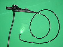
Gastrointestinal lavage
The endoscopically guided segmental gastrointestinal lavage (short intestinal lavage ; English Endoscopically guided segmental lavage ) (from Latin gaster 'stomach', intestinum 'intestine' and lavare 'washing') is a medical diagnostic procedure to detect a suspected food allergy in the stomach To determine locally formed immunoglobulins against food components in the intestinal tract, the detection of which in conventional allergy tests ( prick test or serum IgE ) is not always possible.
Test principle
Antibodies against food components come in a small amount in nearly all immune active regions of the digestive tract (especially in the terminal ileum , reactive and rectum (here especially the recto-sigmoid transition) before. A quantitative increase can to an existing food allergy point. In particular, the detection An increased concentration of antibodies of the IgE class has proven to be a meaningful indicator. However, it is more time-consuming in food allergy sufferers than, for example, in those allergic to insect venom , since in food allergies a significant IgE serum level can only be detected in the blood after long exposure and sensitization In spite of the food allergy in the serum, no increased IgE level can be detected, a local or sero-negative gastrointestinal food allergy may still be present.
The aim of intestinal lavage is to detect the antibodies that are formed by the intestinal mucosa (e.g. intestinal mucosa ) during an endoscopic examination directly on site (locally) and thus in the greatest possible concentration using the ImmunoCAP procedure. The challenge lies in measuring the immunoglobulins in the lavage fluid, the concentration of which can be below that of a normal serum test. In the subsequent analysis one tries to detect the concentration of the intestinal specific IgE antibody (e.g. against soy, cow's milk, nuts etc.) in order to determine the food components to which the body is sensitized. If there is an increased degree of sensitization (intestinal specific IgE> 0.35 U / mg protein) with a suspected food allergy, the patient can become symptom-free by avoiding those foods or he must be subjected to a provocation test with these allergens.
Test execution
Intestinal lavage is performed as part of a gastroscopy or colonoscopy ( enteroscopy or colonoscopy ). It is usually part of a detailed functional tissue diagnosis and supplements the biopsy diagnosis and the usual gastroenterological differential diagnosis.
After reaching the desired section of the intestine, about 50-100 ml of 0.9% saline solution is injected into a section of the intestine through the working channel of the endoscope . The fluid remains there for a specified period of time (one minute) and absorbs the secreted antibodies (IgE) and immune mediators ( ECP , trypsin , TNF and others) from the intestinal mucosa. The liquid is then sucked off and collected. Mixing with protease inhibitors prevents enzymatic breakdown of the antibodies.
In addition, biopsies are taken for each section of the intestine during endoscopy , which provide information about histamine , ECP , DAO and tryptase values or mast cell and eosinophil activity.
The lavage liquid is then frozen at −80 ° C, while the IgE antibodies remain stable. Biopsies removed are (in contrast to the usual pathology biopsies) not placed in formalin, but immediately after removal are quick- frozen in liquid nitrogen or used alive for allergy diagnostics ( see also mucosa oxygenation ). The samples preserved in this way can then be sent to a laboratory for evaluation.
| examination | 1. Localization | 2. Localization | 3. Localization |
|---|---|---|---|
| Oral lavage | Oral cavity | - | - |
| Lavage during gastrojejunoscopy | Duodenum | middle jejunum | - |
| Lavage during colonoscopy | terminal ileum | appendix | Recto - sigmoid junction |
Sample preparation
A number of laboratory values that are important for allergy diagnostics can be determined directly from the lavage fluid. These include the eosinophilic cationic protein (ECP), mast cell tryptase , TNF-a and total protein. The detection of specific antibodies against the food allergens (nuts, soy, wheat and rye flour, meat, poultry etc.) is only possible after the lavage liquid has been concentrated. It is then examined for total and specific IgE concentrations in an ImmunoCAP device , similar to the serum IgE determination . It is based on the ELISA principle: allergens bound on spongy disks (CAPs) are specifically recognized and bound by the IgEs. The remaining antibodies are removed by rinsing. Then fluorescence- labeled antibodies are added, which bind to the Fc region of the other IgEs. Unnecessary antibodies are washed away again. If you now irradiate the antibodies with light of a certain wavelength , they emit light in a slightly larger wavelength. This emitted wavelength can be measured in a targeted manner and the IgE quantity can thus be determined quantitatively.
Expressiveness
Oral, gastrointestinal or endoscopically controlled lavage is based on the same principle as a serum IgE determination . It is based on the fact that an allergy of the immediate type is preceded by IgE formation. However, the amount of IgE (especially if you avoid foods to which you are allergic) can be very small and / or not detectable in the serum. If the local IgE formation is present in the intestine (IgE-positive plasma cells), the specific IgE antibodies can possibly be detected there in a higher concentration (seronegative gastrointestinal-mediated allergy). Similar immune findings also exist in celiac disease (local transglutaminase IgA antibodies). A positive result (IgE level> 0.35 U / mg protein) does not necessarily mean that there is an allergy. The most meaningful evidence of the presence of a food allergy is found when reproducible symptoms occur during the provocation with this food and / or when the symptoms, the respective illness and / or complication, often associated with one, during abstinence (complete avoidance of the food) Decrease in IgE levels, no longer occur. When examined in isolation from studies of a selected study population, the sensitivity of the local IgE determination from the lavage sensitivity (correct exclusion of a food allergy) is 91% and the specificity (correct exclusion of a food allergy) 75%. If you also look at the ECP level, which is also obtained via the lavage, you reach a sensitivity of 91 percent and a specificity of 80 percent. The endoscopically guided segmental gastrointestinal lavage is an endoscopic method for the detection of food allergies; the double-blind placebo-controlled food challenge continues to be the gold standard , although it is time-consuming and carries the risk of an anaphylactic reaction .
Specific IgE antibodies
There are a large number of different antigens for the detection of allergen-specific IgE on the market. The orally ingested can be divided into:
| category | Examples |
|---|---|
| Medicines (partly also iv) | Antibiotics (amoxycilloyl, ampicilloyl, cefaclor, penicilloyl G&V), chlorhexidine, chymopapain, gelatine, insulin (human, beef), morphine, pholcodine, protamine, suxamethonium, tetanus toxoid |
| Food - Cereals / Flours | Buckwheat flour, spelled, barley flour, gluten, oat flour, millet, linseed, lupia seeds, corn flour, rice, rye flour, sesame meal, wheat flour |
| Food - fish | Eel, cichlid, trout, flounder, herring, cod, carp, salmon, mackerel, anchovy, sardine, plaice, hake, saithe, sole, tuna, clam, catfish, grouper, pikeperch |
| Food - Meats | Moose meat, mutton, chicken, rabbit meat, horse meat, beef, pork, turkey meat |
| Food - Chicken Egg | Conalbumin, egg yolk, egg white, ovalbumin |
| Food - vegetables | Avocado, bamboo shoots, cauliflower, bean, broccoli, mushroom, pea, fennel, cucumber, carrot, potato, chickpea, garlic, cabbage, pumpkin, lentil, olive, Brussels sprouts, beetroot, lettuce, celery, soybean, asparagus, spinach, sweet potato , Tomato, onion |
| Food - Spices | Anise, basil, chilli pepper, curry, dill, clove, ginger, caraway, bay leaf, mint, nutmeg, oregano, paprika, parsley, pepper, sage, thyme, vanilla, cinnamon |
| Food - Milk / Dairy Products | Cheddar cheese, milk protein, alpha-lactalbumin, beta-lactoglobulin, casein, lactoferrin, (cow, sheep, mare, goat) milk, whey, blue cheese |
| Food - Clams / Shellfish | Oysters, crayfish, shrimp, lobster, scallop, crab, lobster, mussel, octopus, snail |
| Food additives | Guar Kernel (E412), Gum arabic (E414), Carob (E410), Carmine Red (E120), Targant (E413) |
| Food - nuts | Cashew nut, peanut, sweet chestnut, hazelnut, coconut, almond, Brazil nut, pecan nut, pine nut, pistachio, walnut |
| Food - fruit | Pineapple, apple, apricot, banana, pear, blueberry, blackberry, date, strawberry, fig, grapefruit, rose hip, raspberry, currant, kaki fruit, cherry, kiwi, lime, lychee, mandarin, mango, orange, papaya, passion fruit, peach, Plum, lingonberry, star fruit, watermelon, grape, lemon |
| Food - Other | Baker's yeast, fennel seeds, honey, hops, coffee, cocoa, pumpkin seeds, malt, poppy seeds, rapeseeds, mustard, tea, sugar beet seeds |
Source:
Since the costs for the individual IgE determinations or mediator tests are high (e.g. GOÄ 14.57 €), a selection of five to 15 allergen tests (mostly staple foods) is made, depending on the anamnesis, clinic, and others Found. The most common allergens in food allergies:
| Children / adolescents | Adults |
|---|---|
| Cow's milk, milk components, chicken eggs, wheat, nuts, soy products, fruits, mold products, vegetables, grains, meat, fish | Nuts, soy products, celery, pollen-associated foods (e.g. fruit), wheat, rye, vegetables, oats, milk components, fish, seafood, meat, mold products, cow's milk, hen's eggs, poultry |
Source:
While both private and statutory health insurance companies cover the costs of determining ten different specific IgEs in the serum, so far only private patients have been paid for IgE determination using intestinal lavage.
Since even the smallest amounts of antigens can trigger an allergy, it is worthwhile in exceptional cases to test for potential allergens that are normally not ingested orally (e.g. inhaled allergens, pollen, mold; e.g. eosinophilic esophagogastroenteritis ).
Risks and Complications
These are limited to the general risks of a colonoscopy.
History and Development
1906: Clemens von Pirquet describes allergies
1940: Irving Gray provides the scientific proof of food allergies: IgE release in the mucosa of the digestive tract
Despite the progress in research, it has not been possible to develop a satisfactory detection method for a long time. Neither the prick test nor the IgE detection in the serum show reliable results in food allergies; they are productive in individual subpopulations (e.g. atopic eczema , oral allergy syndrome), but not in all patient collectives. The IgE level in the blood is sometimes unreliable or too low to rule out a food allergy. A study shows: 20% of the test persons who had previously been certified to have a food allergy by means of double-blind placebo-controlled oral food challenge (DBPCFC) did not show any increased IgE values in the blood serum ( sero-negative gastrointestinal-mediated allergy )
1985: Svein Kolmannskog tries a food allergy test using IgE detection in stool.
1996: First use of an intestinal lavage on a patient for whom the usual diagnostics with prick and serum IgE tests remained inconclusive despite severe anaphylactic reactions. In her, locally formed IgE antibodies against pork could be detected in different intestinal sections of the upper and lower gastrointestinal tract. The subsequent review of the lavage findings by DBPCFC in the intensive care unit led to abdominal pain, flushing and a drop in blood pressure. When the allergy was reproduced by repeated oral provocation 10 years after the parental leave measures, it was shown that the pork allergy continued to cause symptoms and that the symptoms were still triggered in a weakened form. Likewise, in certain patient subpopulations with z. B. Irritable bowel syndrome (IBS), inflammatory bowel disease (IBD), microscopic colitis, gastrointestinal bleeding as a result of allergies, mastocytosis or eosinophilic diseases, allergen identification with clear symptom improvement or healing of the disease can be achieved (see also Fig. Diseases with potential allergy and immune phenomena) . However, such tangible immune phenomena are not found in all persons with the above-mentioned diseases, but only in certain subgroups, so that a precise and standardized preliminary diagnosis is clinically necessary before performing this special gastroenterological immunodiagnosis.
literature
- M. Raithel, EG Hahn, H.-W. Baenkler: Clinic and diagnostics of food allergies: Gastrointestinal mediated allergies grade I to IV. In: Deutsches Arzteblatt. 2002; 99 (12): A-780 / B-641 / C-599.
- Y. Zopf, H.-W. Baenkler, A. Silbermann, EG Hahn, M. Raithel: Differential diagnosis of food intolerance. In: Deutsches Arzteblatt International. 2009; 106 (21), pp. 359-369.
- A. Nabe: Determination of tumor necrosis factor alpha, total and specific IgE in food allergy sufferers and chronic inflammatory bowel diseases in the lower gastrointestinal tract by means of endoscopically controlled segmental intestinal lavage. (PDF; 659 kB) Dissertation. Univ. Erlangen-Nuremberg 2009.
Individual evidence
UK Erlangen: Clinical presentation, rational diagnostics and therapy guidelines presentation (PDF; 1.1 MB)
- ↑ M. Raithel, EG Hahn: Functional diagnostic tests to objectify gastrointestinally mediated forms of allergy. In: Allergology. 1998; 21/2, pp. 51-64.
- ↑ a b c D. Schwab, M. Raithel, P. Klein, S. Winterkamp, M. Weidenhiller, M. Radespiel-Troeger, J. Hochberger, EG Hahn: Immunoglobulin E and eosinophilic cationic protein in segmental lavage fluid of the small and large bowel identify patients with food allergy. In: American Journal of Gastroenterology . Volume 96, Issue 2, February 2001, PMID 11232698 , pp. 508-514.
- ↑ a b c d M. Raithel et al.: Allergic gastrointestinal diseases (gastrointestinal mediated allergies I-IV °): From concept to reality. In: Journal for Environmental Medicine. No. 8 (2000), pp. 355-365. (Article) ( Page no longer available , search in web archives ) Info: The link was automatically marked as defective. Please check the link according to the instructions and then remove this notice.
- ^ U. Bengtsson: IgE-positive duodenal mast cells in patients with food-related diarrhea. In: Int Arch Allergy Appl Immunol. 1991; 95 (1), pp. 86-91, PMID 1917114 .
- ↑ Raithel / Herold Innere Medizin 2010, p. 453.
- ↑ Phadia; 'ImmunoCAP'; (Article) (PDF; 1.2 MB)
- ↑ a b M. Raithel et al.: Clinic and diagnosis of food allergies: Gastrointestinal mediated allergies grade I to IV. In: Dtsch Ärzteblatt 2002. No. 12 (2002), pp. 780–786. (Items)
- ↑ a b c Zopf, Yurdagül and others: Differential diagnosis of food intolerance . In: Dtsch Arztebl Int . No. 106 (21) , 2009, pp. 359-370 ( (Article) ).
- ↑ L. Söderström, A. Kober, S. Ahlstedt, H. de Groot, C.-E. Lange, R. Paganelli, MHWM Roovers, J. Sastre: A further evaluation of the clinical use of specific IgE antibody testing in allergic diseases. In: Allergy. Volume 58, 9th edition. September 2003, PMID 12911422 , pp. 921-928.
- ^ Eckhard Schönau, Emil G. Naumann, Alfred Längler, Josef Beuth : Pediatrics integrative: Conventional and complementary therapy. Elsevier, Urban and Fischer, Munich 2005, ISBN 3-437-56500-1 , p. 241, Googlebooks
- ↑ Phadia (article) ( page no longer available , search in web archives ) Info: The link was automatically marked as defective. Please check the link according to the instructions and then remove this notice. (PDF; 1.2 MB)
- ↑ VDGH: EBM & GOÄ excerpt
- ↑ B. Rodeck, K.-P Zimmer: Pediatric gastroenterology, hepatology and nutrition. 1st edition. Springer, Heidelberg 2008, ISBN 978-3-540-73968-5 , pp. 222-230, (article)
- ^ Gray et al .: Studies in mucous membrane hypersensitivity. In: Ann. Int. Med. 13: 2050, May 1940, (Article)
- ↑ M. Walzer: Mechanism of Allergy. PMID 19312158 .
- ↑ K. Aas et al .: Standardization of allergen extracts with appropriate methods. The combined use of skin prick testing and radio-allergosorbent tests. In: Allergy . 1978 Jun; 33 (3), PMID 81622 , pp. 130-137.
- ^ U. Bengtsson: Double blind, placebo controlled food reactions do not correlate to IgE allergy in the diagnosis of staple food related gastrointestinal symptoms. In: Good . 1996 July; 39 (1), PMID 8881824 , pp. 130-135. (Items)
- ^ S. Kolmannskog: Immunoglobulin E in feces from children with allergy. Evidence of local production of IgE in the gut. PMID 3967940 .
- ↑ K. Sasai et al .: IgE levels in faecal extracts of patients with food allergy. In: Allergy. 1992; 47, PMID 1285567 , pp. 594-598.
- ↑ M. Raithel, S. Winterkamp, M. Weidenhiller, S. Müller, EG Hahn: Combination therapy using fexofenadine, disodium cromoglycate, and a hypoallergenic amino acid - based formula induced remission in a patient with steroid - dependent, chronically active ulcerative colitis . In: International Journal of Colorectal Disease 2007 22 (7), pp. 833-839.
- ↑ M. Weidenhiller, S. Müller, D. Schwab, EG Hahn, M. Raithel, S. Winterkamp: Microscopic (collagenous and lymphocytic) colitis triggered by food allergy. In: Gut 2005. 54, pp. 312-313.


